
Figure 5.
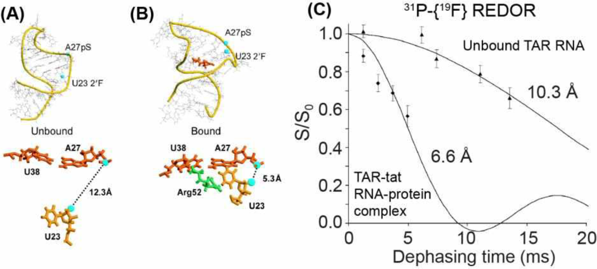
31P-19F REDOR for distance measurements in the tat-TAR complex56. (A) Solution NMR model of TAR RNA (PDB 1ANR), showing locations of the pS-tagged A27 and the fluorodeoxyuridine at U23. (B) Model of tat-bound TAR RNA (PDB 1ANJ), showing a decrease of the 31P-19F distance. The structural model shows that binding of R52 in tat changes the RNA conformation. (C) 31P-19F REDOR dephasing indicates that tat binding shortened the distance between pS-A27 and fluoro-U2356.