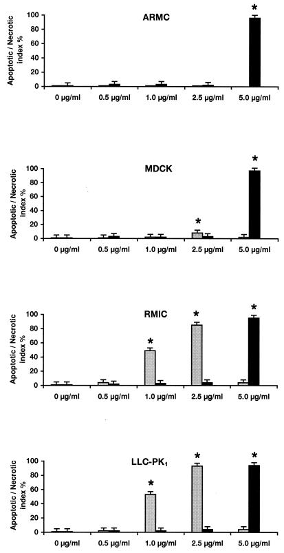
FIG. 1.
Determination of the apoptotic (gray columns) and necrotic (black columns) index values in renal tissue culture cell lines after application of AmB. Confluent monolayers of rat mesangial cells (ARMC), canine distal tubular cells (MDCK), rat medullary interstitial cells (RMIC), and porcine proximal tubular cells (LLC-PK1) were exposed to various AmB concentrations. Apoptotic cells were detected immunohistochemically by end labeling of DNA strand breaks (TUNEL assay) or necrotic cells by the trypan blue exclusion assay, respectively. Each set of experiments was repeated three times. The columns represent the means of results from all four experiments. Standard deviations of the means are indicated by error bars. ∗, Value differs significantly from that of untreated control groups (P < 0.05).

