
FIGURE 3.
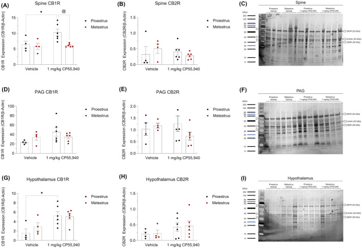
Expression of cannabinoid receptors in brain regions underlying cannabinoid‐induced thermal antinociception and hypothermia. SDS‐PAGE and immunoblotting were performed on (A–C) spine, (D–F) PAG, and (G–I) hypothalamus tissue to quantify their optical density of the cannabinoid receptors. (A, D, G) CB1R expression of region‐specific experimental groups. (B, E, H) CB2R expression of region‐specific experimental groups. (C, F, I) Representative images of the region‐specific CB1R, CB2R, and β‐Actin bands identified using a ladder. (A–I) β‐Actin was used as the housekeeping gene to normalize all cannabinoid receptor densities. Data is reported as ratios of adjusted total band volumes of optical densities. Each point on the graphs represent individual mice or n. Bars on the graphs illustrate means ± SEM, where n = 4 (vehicle) and n = 6 (1 mg/kg CP55,940). All means were compared between 1 mg/kg CP55,940 and vehicle groups within estrus cycle phases, as well as between proestrus and metestrus groups within treatments. A two‐way ANOVA followed by Tukey's post hoc analyses were used to determine significance. *p < .05 compared with vehicle within estrus cycle phase. @ p < .05 compared with proestrus within treatment