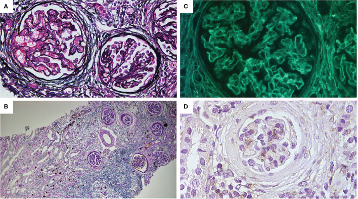
Figure 3.
(A) Mild thickening of the capillary wall and glomerular basement membrane. Bowman capsule enlargement and reduplication (periodic Schiff-methenamine silver, original magnification: ×400). (B) Interstitial and peri-tubular IgG4 deposits. Mild tubular atrophy and interstitial fibrosis. Moderate focal and periglomerular lymphoplasmacytic infiltrate (PAS + IgG4 IHC, original magnification: ×100). (C) Glomerular sub-epithelial deposits with granular pattern (immunofluorescence for anti-PLA2R antibodies, original magnification: ×400). (D) Absence for THSD7A glomerular expression (IHC original magnification: ×200).

