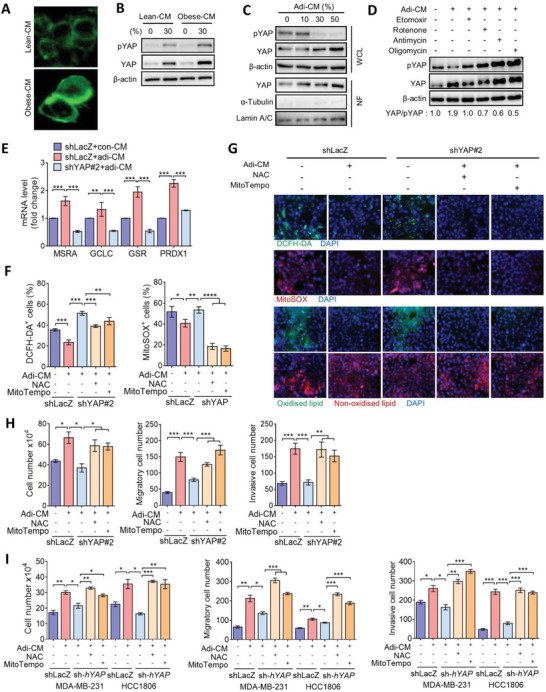
Figure 6.

Adipocytes promote aggressiveness of TNBC in a YAP‐driven antioxidant manner. A) BODIPY staining of lipid accumulation in Py8119 cells incubated with lean‐CM or obese‐CM for 24 h. B) Western blot analysis of total and phosphorylated YAP levels in Py8119 cells treated with lean‐CM or obese‐CM. C) Western blot analysis of total and phosphorylated YAP levels in the whole‐cell lysate (WCL) and total YAP levels in the nuclear fraction (NF) of Py8119 cells treated with different concentrations of adi‐CM. D) Py8119 cells were pretreated with etomoxir, rotenone, antimycin, or oligomycin and then incubated with adi‐CM for 24 h. Expressions of YAP and phosphorylated YAP were assessed by Western blotting. E) qPCR analysis of MSRA, GCLC, GSR, and PRDX1 gene expressions in Py8119/shLacZ and shYAP cells exposed to adi‐CM. (F‐H) Py8119/shLacZ and shYAP cells were pretreated with NAC (2 × 10⁻³ m) or MitoTEMPO (5 × 10⁻⁶ m) followed by incubation with adi‐CM for another 24 h, F) DCFH‐DA+ and MitoSOX+ cells were analyzed by flow cytometry, G) fluorescent microscopic observations of DCFH‐DA, MitoSOX, and BODIPY 581/591 C11 staining, and H) cell proliferation and transwell migration and invasion assays were performed. I) MDA‐MB‐231/shLacZ, HCC1806/shLacZ, MDA‐MB‐231/shYAP, and HCC1806/shYAP cells were pretreated with NAC or MitoTEMPO followed by incubation with adi‐CM for 24 h. Cell proliferation and transwell migration and invasion assays were performed. Data are presented as the mean ± SD. * p < 0.05, ** p < 0.01, *** p < 0.001, as determined by an unpaired two‐tailed Student's t‐test.
