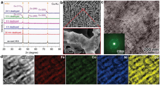
Figure 2.

a) XRD patterns of the dealloyed FeCoNiCu HEA at different etching times. b) SEM characterization of the 8 h dealloyed FeCoNiCu HEA and the enlarged image of the ligaments with secondary mesoporosity. c) TEM images of the secondary spinodal decomposition‐like mesoporous structure. The inset shows the corresponding SAED pattern. d) HAADF‐STEM image and corresponding elemental mapping of the spinodal decomposition‐like mesoporous Cu enriched phase.
