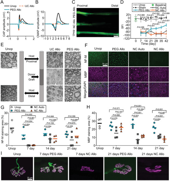
Figure 6.

Neural repair effects of 3D printed NGC. A) Representative compound action potential (CAP) recordings from unoperated control sciatic nerve, PEG‐fused PNA, and negative control PNA. B) Representative compound muscle action potential (CMAP) recordings after stimulating sciatic nerves, recorded from tibialis anterior (TA) muscle. C) Intra‐axonal diffusion evidence for immediate restoration of axonal continuity in PEG‐fused PNA. D) SFI scores show functional recovery over time. E) TEM images showing axons and myelin in cross sections of unoperated control nerve (left) and proximal, graft, and distal segments of negative control PNA (middle) and PEG‐fused PNA (right) at 21 days PO. TEM, transmission electron microscopy. F) Fluorescence images showing cross sections of unoperated control nerve (Unop), PEG‐fused PNA (PEG Allo), NC autograft (NC Auto), and NC PNA (NC Allo) at 7 days PO. G) Time course of % NF‐M staining areas from 7 to 21 days PO. H) Time course of % MBP staining areas from 7 to 21 days PO. I) Confocal images of NMJs immunostained for NF‐M (green) and acetylcholine receptor (AchR, magenta) in animals of unoperated control, PEG‐fused, and negative control PNAs groups at 7th and 21st day PO. Reproduced with permission.[ 89 ] Copyright 2020, Wiley‐VCH.
