
Figure 4.
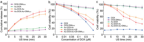
US‐controlled drug release and activation inhibiting cancer cell proliferation. a) The calculated cumulative release of DOX from DNADOX, Au‐DOX, Au‐DOX‐Au dimer, and Au‐DOX‐Au dimer + DNAcom in response to ultrasonication for different times, respectively. The experiments were carried out in triplicate. Mean values: SD from the mean, N = 3 independent experiments. b) Cell proliferation assay involving LNCaP cells with different concentrations of free DOX, DOX+DNADOX, Au‐DOX‐Au dimer without or with 30 min ex situ ultrasonication, and Au‐DOX‐Au dimer + DNAcom with 30 min ex situ ultrasonication, respectively. The experiments were performed in triplicate. Mean values: SD from the mean, N = 3 independent experiments. c) Cell proliferation assay involving LNCaP cells with equivalents of 1 × 10⁻⁶ m free DOX, DOX+DNADOX, Au‐DOX NP, Au‐DOX‐Au dimer, and Au‐DOX‐Au dimer + DNAcom against ex situ ultrasonication for different times, respectively. The experiments were carried out in triplicate. Mean values: SD from the mean, N = 3 independent experiments.