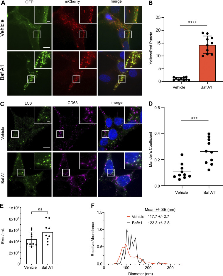
Figure S1.
BafA1 treatment inhibits autophagic flux and modulates EV secretion. (A) Representative images of WT HEK293T cells stably expressing the mCherry-EGFP-LC3 reporter were treated with 20 nM BafA1 or vehicle in serum-free media (BafA1) for 16 h. Scale bar, 10 μm; inset scale bar, 2 μm. (B) Quantification of the ratio of double-positive (mCherry+/GFP+) to mCherry-only (mCherry+/GFP−) LC3 puncta per cell. Statistical significance was calculated by unpaired two-tailed t test (mean ± SEM; vehicle, n = 10; BafA1, n = 10; ****, P < 0.001). (C) Representative images of WT HEK293T cells treated with 20 nM BafA1 or vehicle in serum-free media (BafA1) for 16 h and immunostained for endogenous LC3 and CD63. Scale bar, 10 μm; inset scale bar, 2 μm. (D) A scatter plot of Mander’s coefficients for the co-occurrence of LC3 with CD63 in the immunostained cells in C. Statistical significance was calculated by unpaired two-tailed t-test (mean ± SEM; Vehicle, n = 10; BafA1, n = 10; ***, P < 0.005). (E) Nanoparticle tracking analysis of conditioned media from equal numbers of WT cells treated with vehicle in serum-free media or 20 nM BafA1 (mean ± SEM; n = 3). Statistical significance calculated by unpaired two-tailed t test. (F) EV size distribution from indicated cell treatments in E (mean ± SEM; n = 3).

