
FIGURE 1.
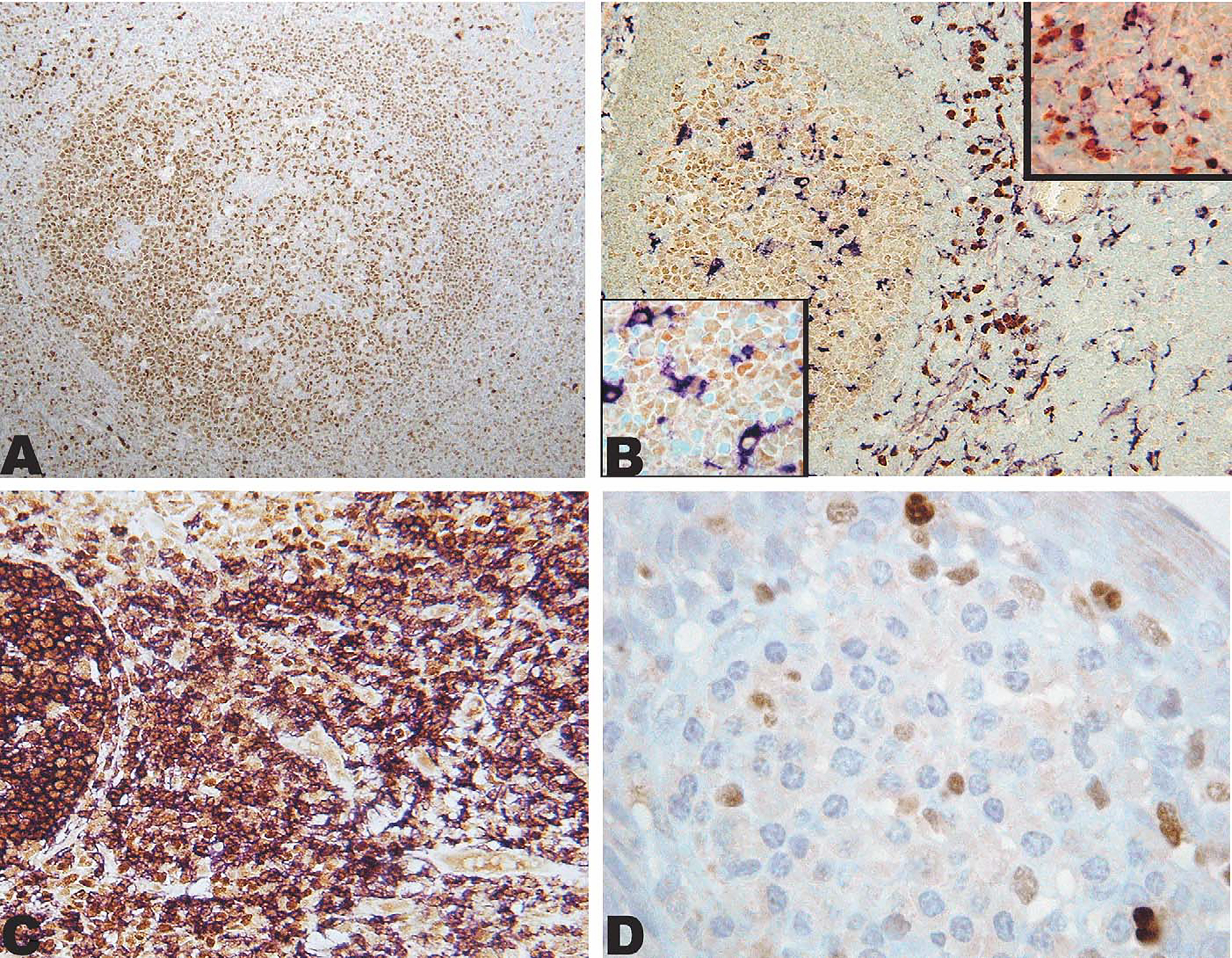
IRF8 expression in reactive lymphoid tissue. A, IRF8 is expressed in a reactive polarized follicle with stronger staining of the germinal center in comparison with the mantle. Strong staining can be seen in scattered interfollicular mononuclear cells. B, Double immunostain showing IRF8 (brown; DAB) expression in CD68 (purple; VIP) positive cells (macrophage/dendritic cells) in the extrafollicular area (upper inset) and weakly in CD68-positive cells (macrophages) in the germinal center (lower inset). C, Double immunostain showing nearly all B cells CD20-positive (purple; VIP) expressing IRF8 (brown; DAB). D, Cluster of plasma cells in the submucosa of a reactive tonsil, rare mononuclear cells are positive with IRF8, whereas plasma cells are negative.