
FIGURE 2.
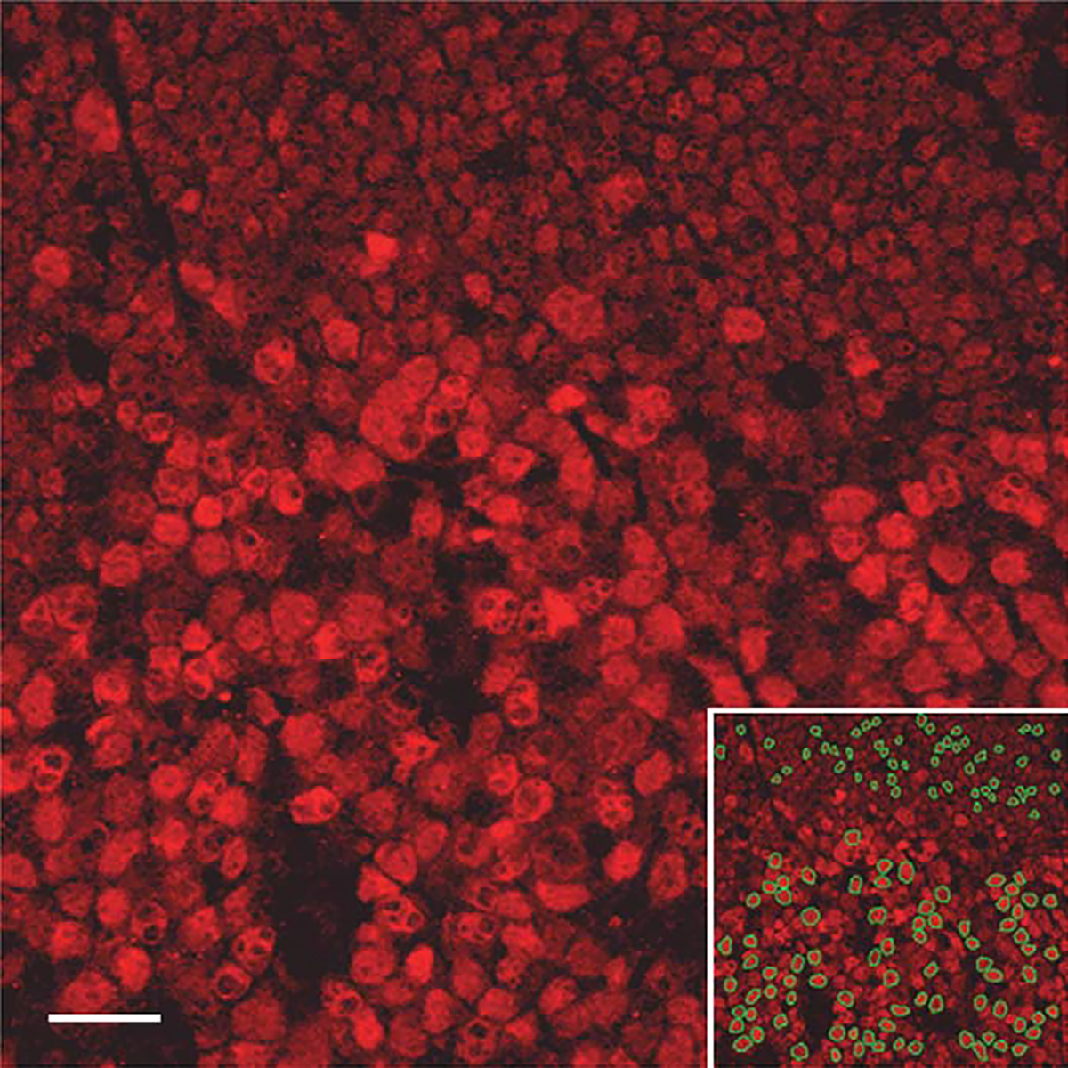
IRF8 expression in follicular B cells by quantitative confocal microscopy: Reactive follicle stained with IRF8 by immunofluorescence (Rhodamine) showing germinal center cells (bottom two-thirds of image) and adjacent mantle cells (top third of image). Lower right inset identifies cells (outlined in green) selected for fluorescence quantitation in each compartment.