
FIGURE 3.
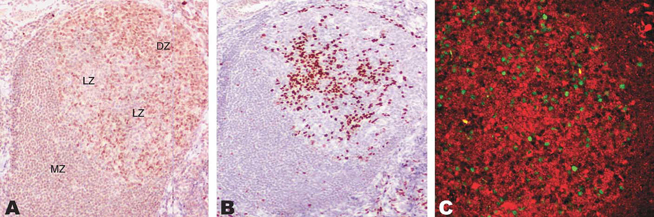
Exclusion between IRF8 and IRF4 in germinal centers shown by sequential immunohistochemical stains and immunoflurorescence. A, The dark zone (DZ) of the germinal center is densely packed by IRF8-positive cells, whereas less numerous positive cells are noted in the light zone (LZ). The mantle zone of the follicle is labeled MZ for reference. B, IRF4-positive cells are present in the light zone of the germinal center. C, Confocal immunofluorescence for IRF8 (red) and IRF4 (green) confirms a mutually exclusive pattern in reactive follicles.