
FIGURE 4.
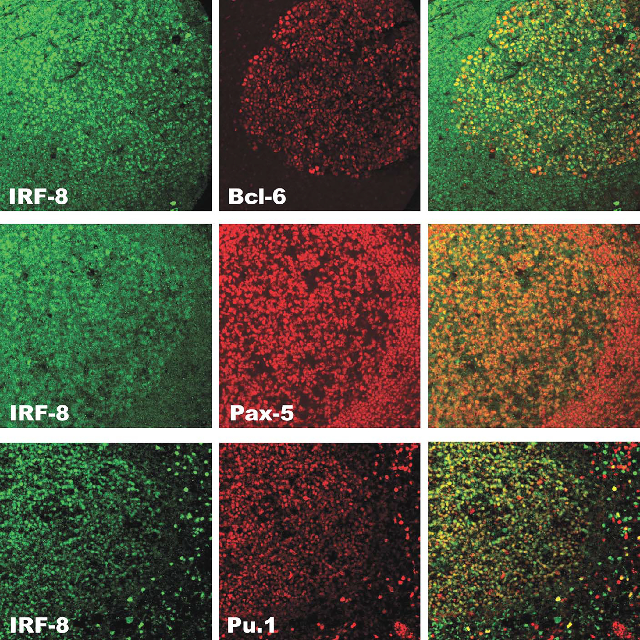
Dual-label confocal immunofluorescence of IRF8 with BCL-6, PAX-5, and Pu.1 in a reactive B-cell follicle. Upper panel. IRF8 (green) is coexpressed with BCL-6 (red) in the germinal center, but not in the mantle as shown in the overlay (yellow-green/yellow-orange). Middle panel. IRF8 (green) is coexpressed with PAX-5 (red) in germinal center and mantle as shown in the overlay (yellow). Lower panel. IRF8 (green) is coexpressed with Pu.1 (red) in the germinal center, mantle and in scattered is interfollicular macrophages (overlay, yellow).