
FIGURE 5.
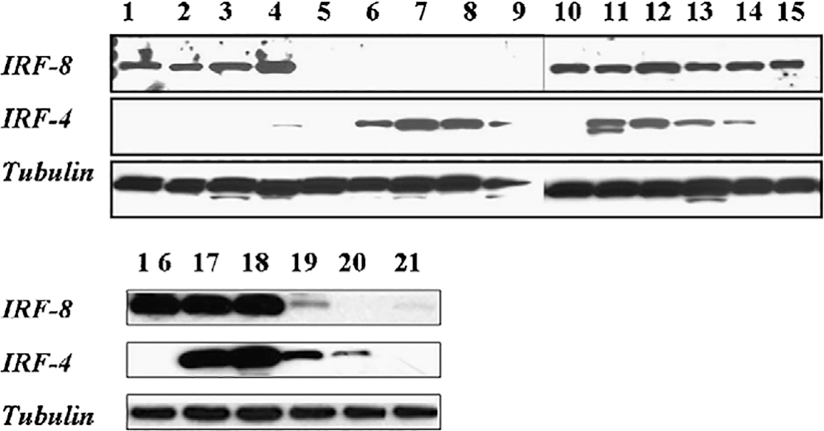
Western blot for IRF8 and IRF4 in cell lines. Western blot analysis of representative cell lines for IRF8 (panel A), IRF4 (panel B), and tubulin as a loading control (panel C). IRF8 is present in DLBCL GCB-like cell lines SUDHL-4, SUDHL-5, SUDHL-6, SUDHL-7 (lanes 1, 2, 3, 16) and negative KMS-12, KMS-20 myeloma (lanes 6, 7), and BC-2, BC-3 primary effusion lymphoma cell lines (lanes 8, 9). In contrast OCI-Ly3 and OCY-Ly10 DLBCL non-GCB–like (lanes 17, 18), Granta and Z-138 MCL (lanes 19, 20), and the JD38 BL cell lines (lane 4) show expression of both IRF8 and IRF4.