
FIGURE 6.
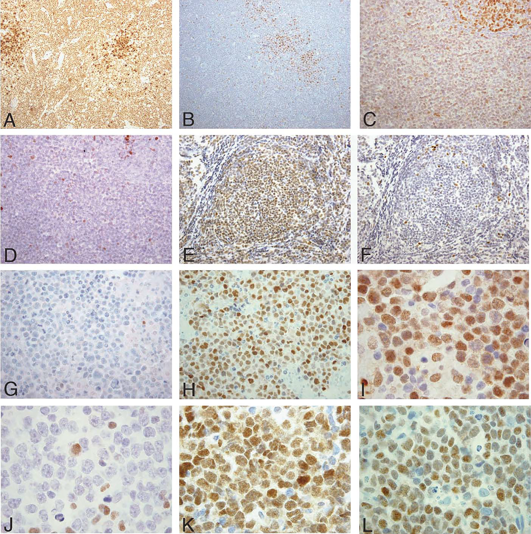
IRF8 and IRF4 expression by immunohistochemistry in B-cell non-Hodgkin lymphomas. A and B, CLL ( × 20 HPF). A, Neoplastic cells express IRF8 with strongest intensity in proliferation centers. B, IRF4 is negative in most cells with the exception of proliferation centers. C and D, MCL ( × 20 HPF). C, IRF8 is weakly expressed in the neoplastic cells. Note the stronger IRF8 expression in a residual reactive germinal center (top right). D, Scattered IRF4-positive cells with the majority of neoplastic cells negative. E and F, FL ( × 20 HPF). E, Neoplastic cells show strong expression for IRF8, and (F) are negative for IRF4. G and H, MM ( × 40 HPF). G, Myeloma cells are negative for IRF8 and (H) positive for IRF4. I and J, DLBCL of GCB type ( × 60 HPF) showing expression of IRF8 (I), but not IRF4 (J). K and L, DLBCL of GCB type ( × 60 HPF) with coexpression of IRF8 (K) and IRF4 (L). [This latter DLBCL was classified as GCB type on the basis on a positive CD10 stain (not shown), according to the criteria of Hans et al11].