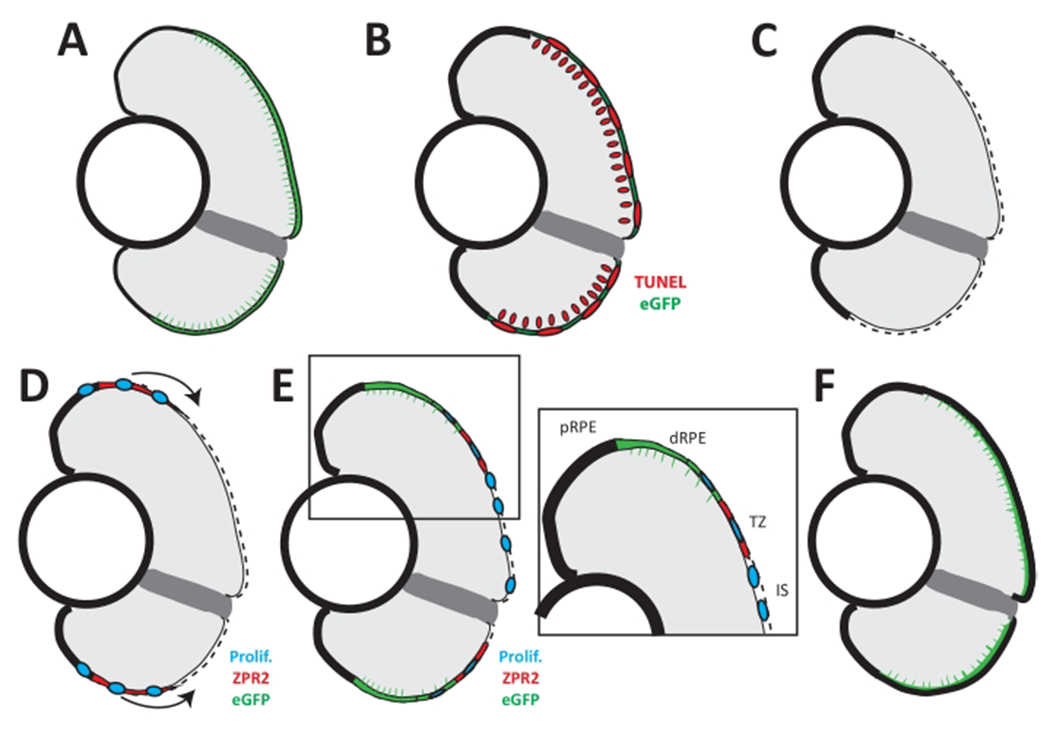
Figure 6: Model of RPE regeneration in larval zebrafish.

(A) nfsB-eGFP is expressed in mature RPE in the central two-thirds of the eye. (B) Application of MTZ leads to apoptosis (TUNEL, red) of RPE and photoreceptors. (C) RPE ablation leads to degeneration of photoreceptors and Bruch’s membrane (dotted line). (D) Unablated RPE in the periphery begin to proliferate and extend into the injury site (blue). (E) As regenerated eGFP+ RPE appear in the periphery, the RPE can be divided into four zones: peripheral RPE (pRPE), differentiated RPE (dRPE), transition zone (TZ), and injury site (IS). (E, inset) Regenerated differentiated RPE (green) appears in the periphery proximal to the unablated peripheral RPE, and contains proliferative cells adjacent to the transition zone. The transition zone consists of still-differentiating RPE cells (ZPR2, red) and proliferative cells (blue). The injury site comprises unpigmented proliferative cells that do not express any RPE differentiation markers. (F) Regeneration of a functional RPE layer and Bruch’s membrane is complete by 14 dpi. This figure and figure legend text have been modified from Hanovice et al. 2019 (Figure 14)3. Please click here to view a larger version of this figure.
