Figure 4.
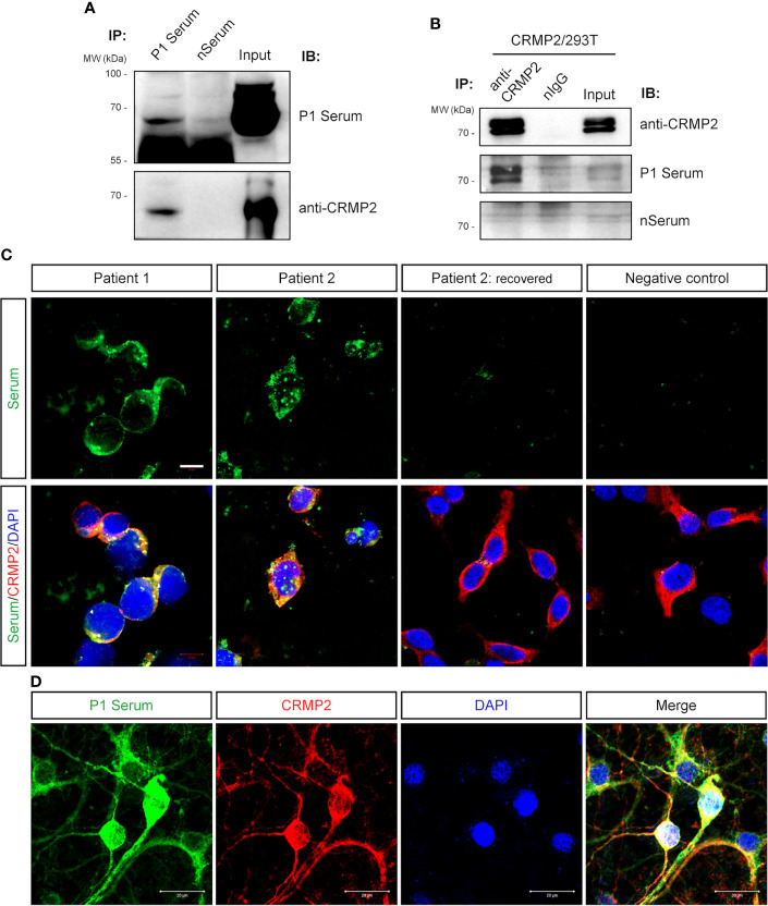
Verification of anti-CRMP2 antibody. (A) IP with P1 patient’s serum was performed with rat brain protein lysate. Immunoblottings were done with P1 patient’s serum (upper panel) and commercial anti-CRMP2 antibody (lower panel). (B) IP with commercial anti-CRMP2 antibody was performed with HEK293T cells overexpressing full-length human CRMP2 (isoform 1). Anti-CRMP2 antibody, P1 patient, or negative control sera were used for Western blotting, separately. (C) CRMP2-expressing HEK293T cells were immunostained with the patients’ sera and anti-CRMP2 antibodies. The sera from the first collection of P1 and P2 patients showed positive staining and co-localized with CRMP2. Serum collected from P2 patient 1.5 years after recovery showed negative staining. (D) Colocalization of P1 patient’s serum and anti-CRMP2 Ab immunostaining in cultured mouse cortical neurons. CRMP2, collapsin response mediator protein 2; DAPI, 6-diamidino-2-phenylindole; IB, immunoblotting; nIgG, normal immunoglobulin G; IP, immunoprecipitation; WB, Western blotting. Scale bars represent 10 μm in (C) and 20 μm in (D).

