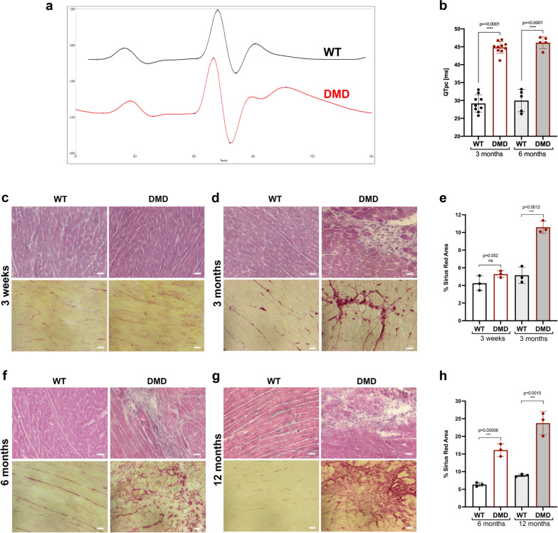
Fig. 4.
Heart functional and histological evaluation in R-DMDdel52 rats. a ECG signal of (black) control and (red) R-DMDdel52 rat at 6 months of age. b Quantification of the QTpc interval in WT and R-DMDdel52 rats at 3 and 6 months of age. c Hematoxylin and eosin (upper panel) and Sirius red (lower panel) staining of heart of 3-week-old WT and R-DMDdel52 rats (scale bar = 50 μm). d Hematoxylin and eosin (upper panel) and Sirius red (lower panel) staining of heart of WT and R-DMDdel52 rats aged 3 months (scale bar = 50 μm). e Quantification of fibrotic area in the hearts of WT and R-DMDdel52 rats aged 3 weeks and 3 months. f Hematoxylin and eosin (upper panel) and Sirius red (lower panel) staining of heart of WT and R-DMDdel52 rats aged 6 months (scale bar = 50 μm). g Hematoxylin and eosin (upper panel) and Sirius red (lower panel) staining of heart WT and R-DMDdel52 rats aged 12 months (scale bar = 50 μm). h Quantification of fibrotic area in the hearts of WT and R-DMDdel52 rats aged 6 and 12 months

