Fig. 5.
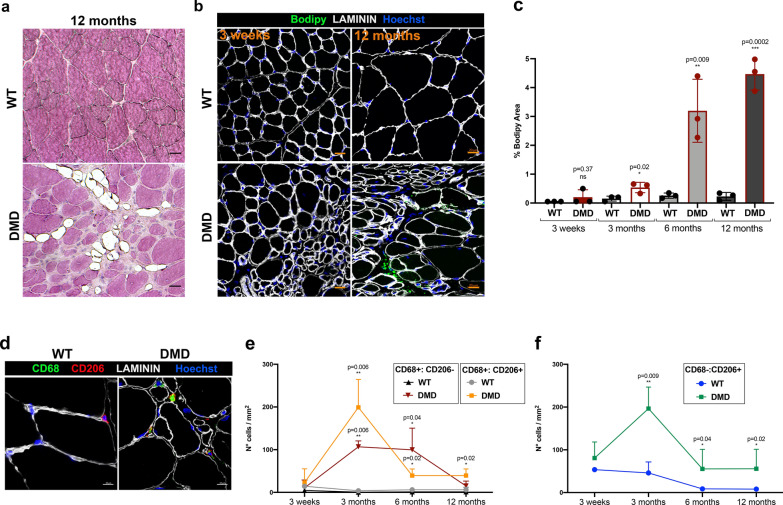
Alterations in fat deposition and inflammatory status in R-DMDdel52 diaphragms. a Hematoxylin and eosin staining of TA from WT and R-DMD-del52 rats aged 12 months (scale bar 20 μm). b Lipid droplet staining by Bodipy immunofluorescence (green) on TA of WT and R-DMDdel52 rats aged 3 weeks and 12 months (scale bar 20 μm). Laminin (white) delineates muscle fibres and nuclei are counterstained with Hoechst (blue). c Quantifications of b. d Immunofluorescence for CD68 (green), CD208 (red), and laminin (white) performed on diaphragms isolated from WT and R-DMDdel52 rats aged 6 months. Nuclei were counterstained with Hoechst (blue). Scale bar 10 μm. e Quantification of the number of CD68-positive or CD206-positive macrophages in 3-week-old and 3-, 6-, 12-month-old WT and R-DMDdel52 muscles. f Quantification of the number of CD68-negative and CD206-positive macrophages (CD206−:CD68+) in 3-week-old and 3-, 6-, 12-month-old WT and R-DMDdel52 muscles

