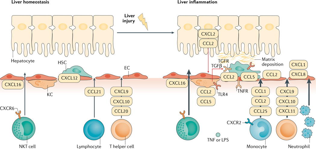
Fig. 1 |. Chemokine pathways in liver homeostasis and inflammation.
In liver homeostasis, Kupffer cells (KCs), liver sinusoidal endothelial cells (ECs) and hepatic stellate cells (HSCs) are in control of surveillance of tissue stress, pathogens and other disturbances. Local immune cells are first responders to chemokines such as CXCL12, maintaining liver homeostasis. When injury escalates, it can cause the release of damage-associated molecular motifs, which activate KCs. This activation induces the production of chemokines and cytokines. Transforming growth factor-β (TGFβ) is produced by KCs and induces the activation of HSCs. CCL2 and CCL5 are important in the process of activation of KCs and HSCs. Tumour necrosis factor (TNF) and lipopolysaccharide (LPS)-induced CXCL1 and CXCL8 are important chemokines that chemoattract neutrophil adhesion and invasion into liver inflammatory sites. Other chemokines, such as CCL1, CCL2 and CCL25, promote the infiltration of monocytes, CXCL9, CXCL10 and CXCL11 promote the infiltration of T helper cells, and CXCL16 promotes the infiltration of natural killer T (NKT) cells. These immune cells also contribute to the local inflammatory milieu. The activation of HSCs by immune cells and chemokines leads to the deposition of collagen in the liver, causing fibrosis. TLR4, Toll-like receptor 4.

