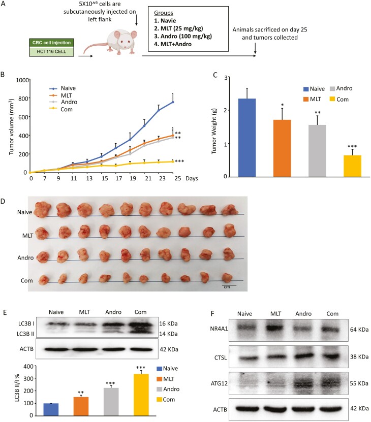
Figure 5.
The combination of MLT and Andro synergistically inhibit xenograft growth in an animal model. (A) Schematic diagram of HCT116 cell-derived xenograft model in nude mice and the treatment schedule of MLT, Andro and their combination. (B) Tumor volume alterations in each treatment group were measured at different time points after inoculation. (C) The measurement of tumor weights within each treatment group 25 days post-inoculation. (D) Representative images of harvested tumors surgically removed from nude mice 25 days post-inoculation. (E) Representative images of LC3B expression in the extracts of xenografts in each treatment group by western blot analysis. The ratios of LC3-II to LC3-I based on the quantification of the bands in the immunoblot were shown in the lower panel. (F) Representative images of NR4A1, ATG12 and CTSL expression in the extracts of xenografts in each treatment group by western blot analysis. Statistical significance: ∗P < 0.05, ∗∗P < 0.01, ∗∗∗P < 0.001.

