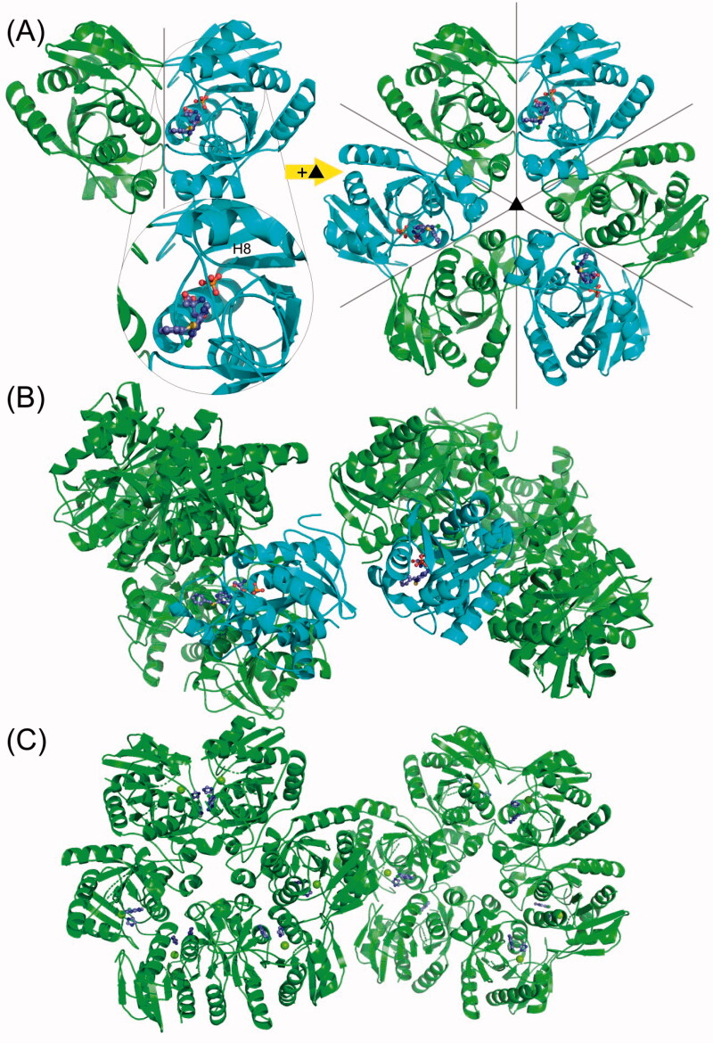
Figure 7.
The overall structure of the H. pylori PNP complexes with 2,6-substituted purines. (A) Complexes with 6BnS-2Cl-Pu and 6BnO-2Cl-Pu crystallise in the cubic space group P 213 with two monomers forming a dimer in the asymmetric unit. One of the monomers has the closed (shown in cyan) and one has the open (shown in green) conformation of the active site. The helix which is segmented to close the active site pocket (see text) is designated H8. The complete hexamer is generated by a crystallographic 3-fold axis, and therefore one hexamer has three open and three closed active sites. Ligands are shown in the ball and stick model. (B) In the crystals of PNP with 6BnS-Pu, two entire hexamers are in the asymmetric unit in the space group P 212121, and each hexamer (shown in cyan) has one closed active site. The closed active sites from different hexamers are next to each other in the crystal packing. (C) Complex of PNP with 2,6-diCl-Pu crystallised also with two hexamers in the asymmetric unit but this time in the P 1 space group. All the monomers are in the open conformation.

