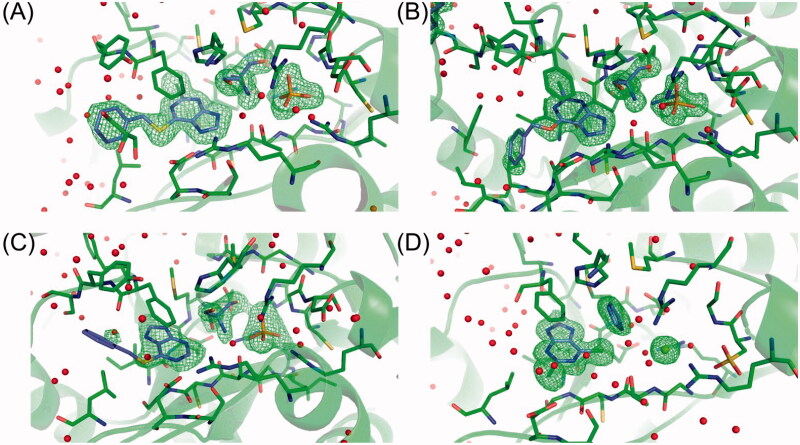
Figure 8.
Comparison of closed active sites in all four structures of H. pylori PNP with (A) 6BnS-2Cl-Pu, (B) 6BnO-2Cl-Pu, (C) 6BnS-Pu, and (D) 2,6-diCl-Pu. C atoms of the ligands are shown in violet, protein C atoms in green, and all other atoms in the CPK colours. The figure shows the electron mFo-DFc difference map density contoured at 3σ level (shown in green). Electron density only around molecules bound in the active sites is shown. Red spheres represent water molecules, and the green sphere on panel (D) – magnesium atom (from crystallisation mother liquor, see text).

