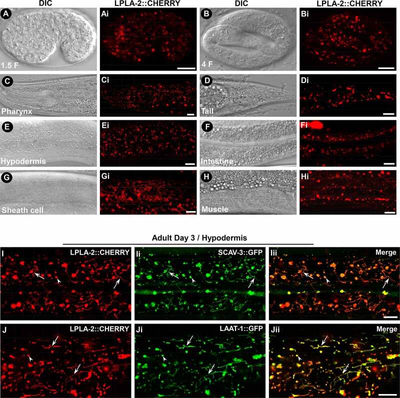
Figure 2.

LPLA-2 is widely distributed and localizes to lysosomes. (A-Hi) DIC and confocal fluorescence images of wild type expressing LPLA-2::CHERRY driven by the lpla-2 promoter. (I-Jii) Confocal fluorescence images of the hypodermis in wild type co-expressing LPLA-2::CHERRY and SCAV-3::GFP (I–Iii) or LAAT-1::GFP (J-Jii). LPLA-2 colocalizes with SCAV-3 and LAAT-1 to both vesicular (arrowheads) and tubular (arrows) lysosomes. Scale bars: 10 µm.
