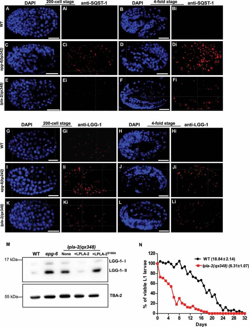
Figure 4.

The autophagy process is partially impaired in lpla-2 mutants. (A-Li) Confocal fluorescence images of wild type (WT), epg-6(bp242) and lpla-2(qx348) embryos at the 200-cell and 4-fold stages stained by anti-SQST-1 (A-Fi) or anti-LGG-1 antibodies (G-Li). DAPI staining shows nuclei in each embryo. (M) Western blot analysis of LGG-1-I and LGG-1-II (lipid-conjugated form) in wild type (WT), epg-6(bp242) and lpla-2(qx348) without and with expression of LPLA-2 or LPLA-2S196A. (N) Quantification of the survival of WT and lpla-2(qx348) L1 larvae in the absence of food. At least 200 animals were scored at each time point in each strain. Three independent experiments were performed and the mean lifespan (mean ± SD) is shown in parenthesis. Scale bars: 10 µm.
