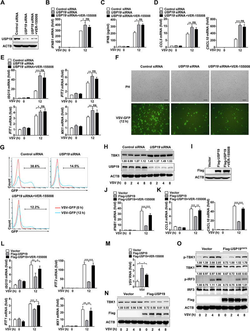
Figure 4.

USP19 negatively regulates VSV-induced IFNB production and the antiviral response in THP1 cells. (A-G) THP1 cells were transfected with control or USP19 siRNA for 48 h, with or without pretreatment with VER-155,008 (5 μM) for 30 min, then USP19 protein levels were analyzed via immunoblotting (A). qPCR analysis of IFNB1 expression for the indicated times after VSV infection (MOI = 1) (B). ELISA analysis of IFNB production in the cell culture supernatants 12 h after VSV infection (C). qPCR analysis of CCL5 and CXCL10 expression 12 h after VSV infection (MOI = 1) (D). qPCR analysis of ISG15, IFIT2, IFIT1, and MX1 expression 12 h after VSV infection (MOI = 1) (E). THP-1 cells were infected with VSV-GFP (MOI = 1) for 12 h and the cells were imaged under a confocal microscope (F). The percentage of GFP+ cells was determined via flow cytometry (G). (H) THP1 cells were transfected with control or USP19 siRNA, and then infected with VSV. Immunoblotting was performed to analyze TBK1 protein levels. (I-M) THP1 cells were transfected with Flag-USP19 plasmids or an empty vector for 24 h, with or without pretreatment with VER-155,008 (5 μM) for 30 min. USP19 expression was analyzed via immunoblotting using a Flag antibody (I). qPCR analysis of IFNB1 (J), CCL5, CXCL10 (K), ISG15, IFIT2, IFIT1, and MX1 (L) expression and VSV replication (M) for the indicated times after VSV infection (MOI = 1). (N) THP1 cells were transfected with Flag-USP19 plasmids or an empty vector, and then infected with VSV. Immunoblotting was performed to analyze TBK1 protein levels. (O) THP1 cells were transfected with Flag-USP19C607S plasmids or an empty vector, and then infected with VSV. Immunoblotting was performed to analyze indicated proteins levels. The data are representative of three independent experiments. Scale bars: 100 μm. Error bars show the means ± SD. *p < 0.05, **p < 0.01, ***p < 0.001 using Student’s t test.
