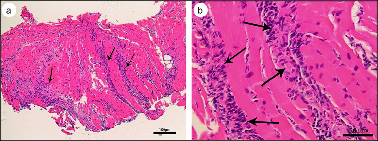
Figure 2.
Myocardial histology, hematoxylin and eosin stain, showing (a) acute myocarditis with predominant neutrophilic inflammation (arrows) (4×) and (b) bands of inflammatory infiltrate invading through the myocardium and consisting predominantly of neutrophils with rare lymphocytes and eosinophils (arrows) (20×).

