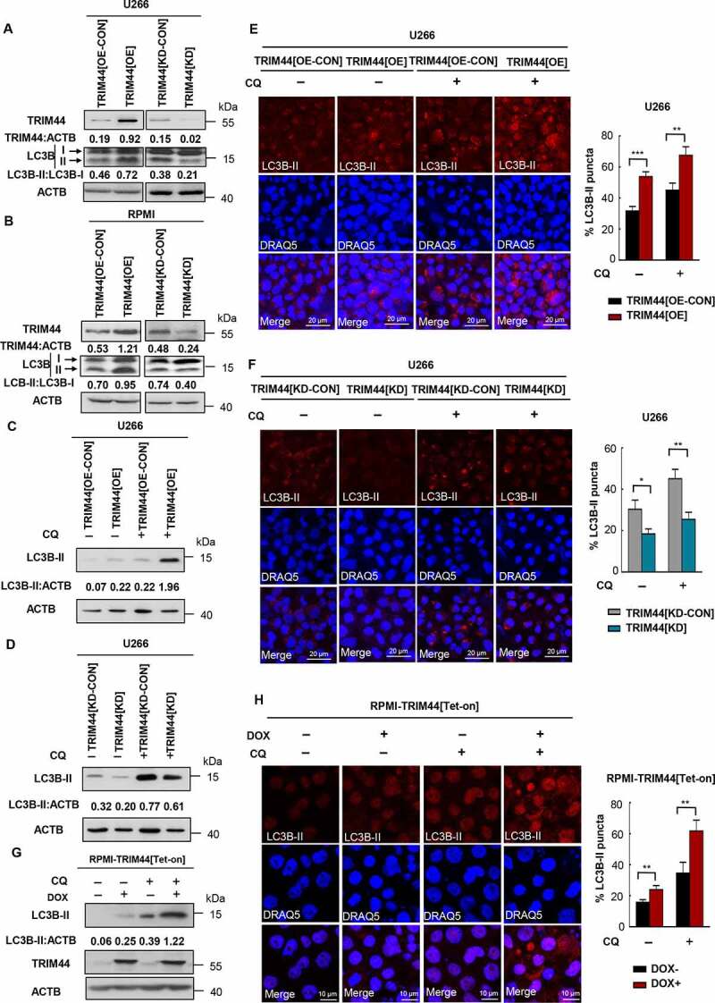
Figure 5.

TRIM44 leads to autophagy activities in MM cells. (a, b) The protein levels of LC3B and SQSTM1 of TRIM44[OE-CON], TRIM44[OE], TRIM44[KD-CON] and TRIM44[KD] MM cells (U266 and RPMI) were assayed by western blots. (c, d) U266 cells (TRIM44[OE-CON], TRIM44[OE], TRIM44[KD-CON] and TRIM44[KD]) were treated with or without CQ (10 μM, 24 h). The protein levels of LC3B were assayed by western blots. (e) TRIM44[OE-CON] and TRIM44[OE] MM cells (U266) were treated with or without chloroquine CQ (10 μM, 24 h). And then cells were fixed and assayed for the appearance of autophagosomes by confocal microscopy. LC3B puncta were analyzed by LC3B-II antibody (magnification is 10 × 60 oil). Scale bars: 20 μm. Quantification of autophagosome formation. Cells with eight or more LC3B puncta were considered to have accumulated autophagosomes. Data are presented as mean % LC3B-positive cells ± SEM in three independent experiments. In each treatment at least 100 cells were analyzed (*, P < 0.05; **, P < 0.01; ***, P < 0.001). (f) TRIM44[KD-CON] and TRIM44[KD] MM cells (U266) were treated with or without CQ (10 μM, 24 h). And then cells were fixed and assayed for the appearance of autophagosomes by confocal microscopy. LC3B puncta were analyzed by LC3B-II antibody (magnification is 10 × 60 oil). Scale bars: 20 μm. Quantification of autophagosome formation. Cells with eight or more LC3B puncta were considered to have accumulated autophagosomes. Data are presented as mean % LC3B-positive cells ± SEM in three independent experiments. In each treatment at least 100 cells were analyzed (*, P < 0.05; **, P < 0.01; ***, P < 0.001). (g) RPMI-TRIM44[Tet-on] cells were treated with or without DOX (1 µg/mL) together CQ (10 μM, 24 h). The protein levels of LC3B were assayed by western blots. (h) RPMI-TRIM44[Tet-on] cells were treated with or without DOX (1 µg/mL) together with CQ (10 μM, 24 h). And then cells were fixed and assayed for the appearance of autophagosomes by confocal microscopy. LC3B puncta were analyzed by LC3B-II antibody (magnification is 10 × 60 oil). Scale bars: 20 μm. Quantification of autophagosome formation. Cells with eight or more LC3B puncta were considered to have accumulated autophagosomes. Data are presented as mean % LC3B-positive cells ± SEM in three independent experiments. In each treatment at least 100 cells were analyzed (*, P < 0.05; **, P < 0.01; ***, P < 0.001).
