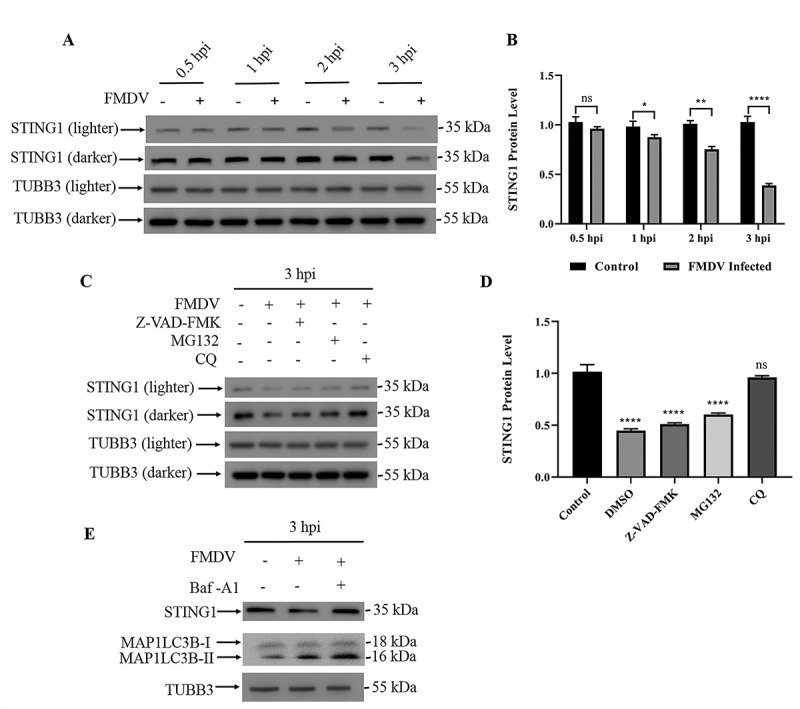
Figure 1.

FMDV infection leads to STING1 degradation through autophagy. (A) Western blot detection of STING1 protein in FMDV-infected PK15 cells (MOI = 1) and uninfected controls (0.5 hpi, 1 hpi, 2 hpi and 3 hpi). TUBB3/β-tubulin was used as a loading control. (B) Gray values of western blot bands in (A) were analyzed by ImageJ, and STING1 values were normalized to TUBB3 intensity. The data show the mean ± SD; n = 3; ns, no significance, *P < 0.1, **P < 0.01, ****P < 0.0001. (C) Western blot detection of STING1 in cells with the indicated treatment. TUBB3 was used as a loading control. PK15 cells were either untreated or treated with the pan-caspase inhibitor Z-VAD-FMK, protease inhibitor MG-132 or autophagy inhibitor CQ when infected with FMDV (MOI = 1). (D) Gray values of western blot bands in (C) were analyzed by ImageJ, and STING1 values were normalized to TUBB3 intensity. The data show the mean ± SD; n = 3; ns, no significance, ****P < 0.0001. (E) Western blot detection of STING1 and MAP1LC3B in PK15 cells infected with FMDV (3 hpi). PK15 cells were treated with or without Baf-A1 when infected with FMDV (MOI = 1). TUBB3 was used as a loading control.
