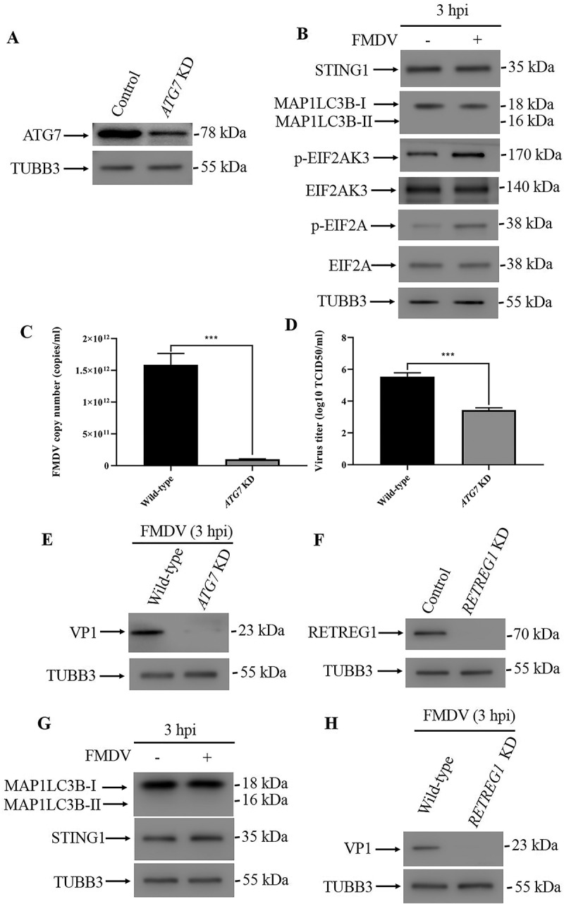
Figure 2.

FMDV infection induced reticulophagy. (A) Stable ATG7 knockdown (KD) PK15 cells were verified by western blot. TUBB3 was used as a loading control. (B) Western blot analysis of STING1, MAP1LC3B, phosphorylated EIF2AK3 and EIF2A levels in FMDV-infected (MOI = 1) and uninfected control cells (3 hpi). TUBB3 was used as a loading control. (C) Copy numbers of FMDV in FMDV-infected wild-type and ATG7 KD cells (MOI = 1, 3 hpi) were measured by qPCR. The data show the mean ± SD; n = 3; ***P < 0.001. (D) Wild-type and ATG7 KD cells were infected with FMDV (MOI = 1), and virus titers were measured by TCID50 (3 hpi). The data show the mean ± SD; n = 3; ***P < 0.001. (E) Western blot analysis of FMDV VP1 protein in FMDV-infected wild-type and ATG7 KD cells (MOI = 1, 3 hpi). (F) Stable RETREG1 KD PK15 cells were verified by western blot. TUBB3 was used as a loading control. (G) Western blot analysis of MAP1LC3B and STING1 in FMDV-infected (MOI = 1) and uninfected RETREG1 KD cells (3 hpi). TUBB3 was used as a loading control. (H) Western blot analysis of FMDV VP1 protein in FMDV-infected wild-type and RETREG1 KD cells (MOI = 1, 3 hpi).
