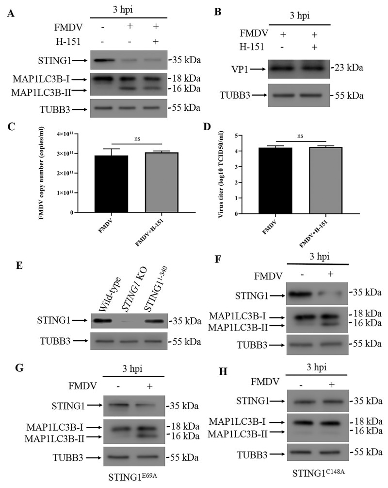
Figure 6.

STING1 activation is not required for autophagy induction. (A) Western blot detection of STING1 and MAP1LC3B in human STING1-reconstituted STING1 KO PK15 cells infected with FMDV (3 hpi). PK15 cells were pre-treated with or without H-151 for 24 h before being infected with FMDV (MOI = 1). TUBB3 was used as a loading control. (B) Western blot detection of FMDV VP1 protein in FMDV-infected human STING1-reconstituted PK15 cells with or without H-151 treatment (3 hpi). (C) qPCR-measured copy numbers of FMDV in FMDV-infected human STING1-reconstituted PK15 cells with or without H-151 treatment (MOI = 1, 3 hpi). The data show the mean ± SD; n = 3; ns, no significance. (D) Human STING1-reconstituted PK15 cells with or without H-151 treatment were infected with FMDV (MOI = 1), and virus titers were measured by TCID50 (3 hpi). The data show the mean ± SD; n = 3; ns, no significance. (E) Stable STING1 KO PK15 cells were transfected with STING1 truncations (residues 1–340) and validated by western blot. (F) Western blot analysis of STING1 and MAP1LC3B in FMDV-infected (MOI = 1) and uninfected PK15 cells that expressed the STING1 truncation (residues 1–340) (3 hpi). TUBB3 was used as a loading control. (G) Western blot analysis of STING1 and MAP1LC3B in FMDV-infected (MOI = 1) and uninfected PK15 cells that expressed the STING1 mutant (STING1E69A) (3 hpi). TUBB3 was used as a loading control. (H) Western blot analysis of STING1 and MAP1LC3B in FMDV-infected (MOI = 1) and uninfected PK15 cells that expressed the STING1 mutant (STING1C148A) (3 hpi). TUBB3 was used as a loading control.
