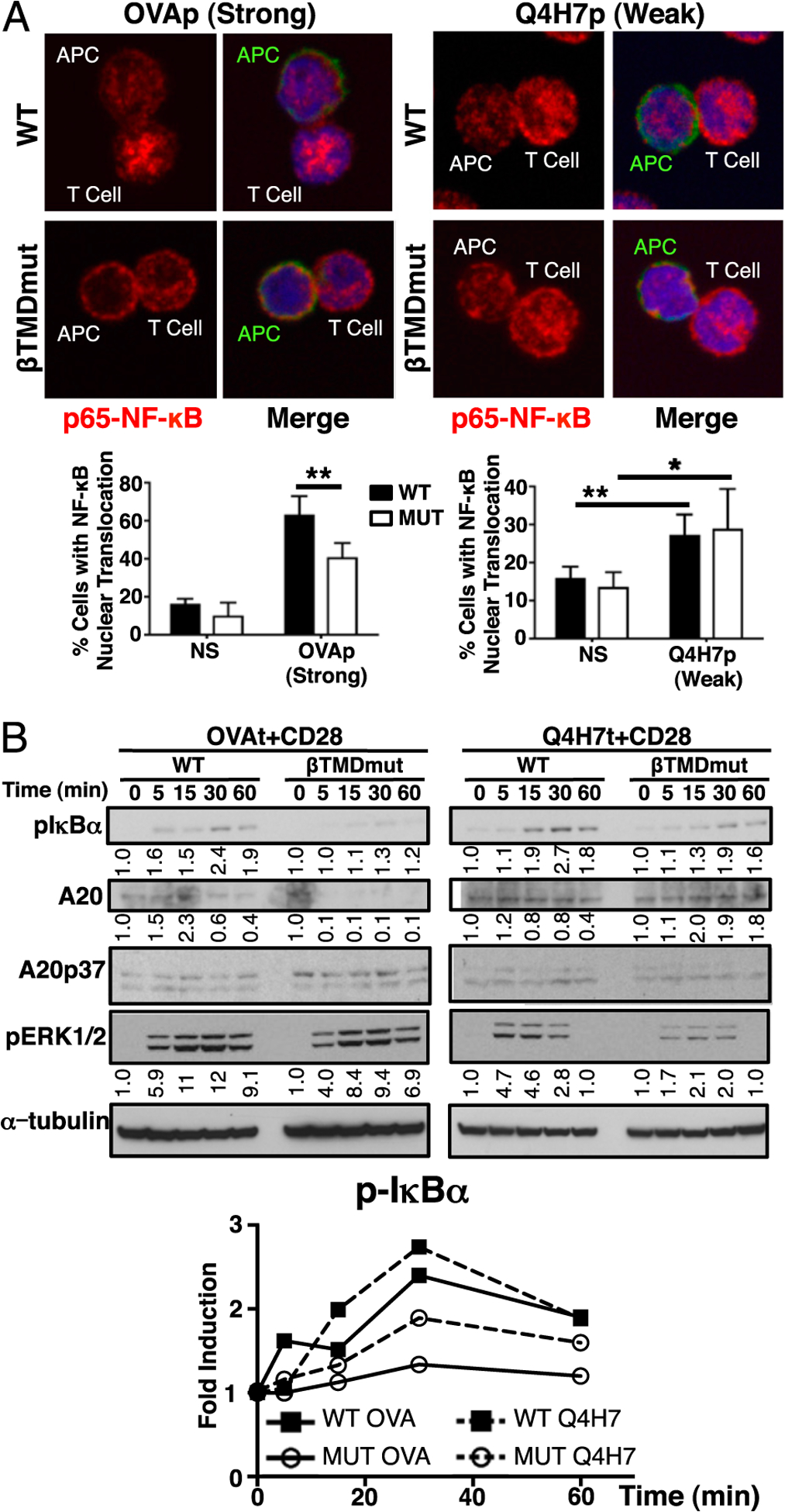
FIGURE 3.

MUT T cells recover NF-κB signaling in response to low-affinity ligands. (A) Nuclear translocation of p65–NF-κB determined by confocal microscopy: CD45.1 (green), NF-κB (red), nucleus (blue). Images are representative of n ≥ 50 conjugates from n ≥ 3 independent experiments. Graphs show the mean percentage ± SD of conjugated T cells that exhibit p65 in the nucleus over unstimulated cell levels. Original magnification ×60. (B) Phosphorylation of IκBα, ERK1/2, and expression of A20 in response to high- and low-affinity TCR ligands was determined in WT or MUT T cells stimulated with Kb-OVA or -Q4H7 tetramers and anti-CD28 Abs by immuno-blot. All immunoblots correspond to the same representative experiment. A representative experiment of n = 4 is shown. Graph and numbers in the blot show relative induction of p-IκBα, A20, p-ERK1/2 levels over unstimulated (time 0), normalized to α-tubulin (loading control). *p ≤ 0.05, **p ≤ 0.005.
