Figure 1.
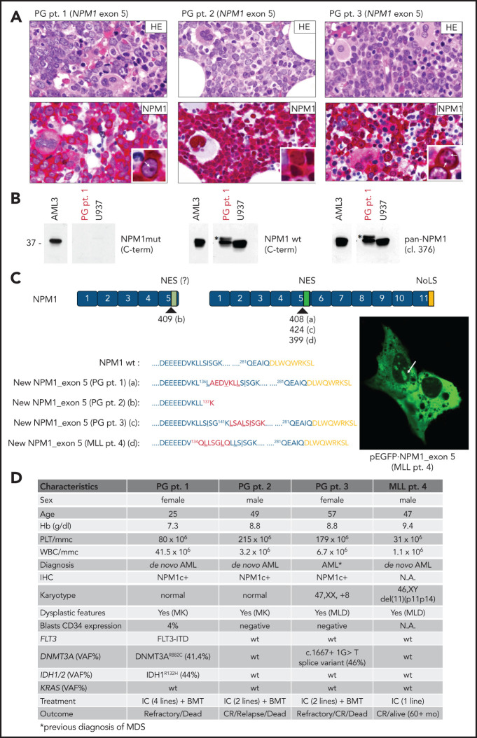
Novel NPM1 gene mutations involving exon 5. (A) IHC staining of BM trephine from PG patients 1, 2, and 3 showing diffuse infiltration by leukemic blasts (hematoxylin and eosin [HE], upper) with aberrant cytoplasmic positivity for NPM1 (NPM1, lower). Besides cytoplamic staining, nucleoli staining is shown in the inset (white arrows) for PG patients 1 and 3. NPM1 staining: mouse monoclonal clone 376 anti-NPM1 N-terminus by antialkaline phosphatase technique with hematoxylin counterstaining. Images were collected using an Olympus B61 microscope with a UPlanApo 40×/0.85 U (40× magnification) and UPlan FI 100×/1.3 NA oil (×100 original magnification) objective for the inset, Camedia 4040 (Dp_soft version 3.2), and Adobe Photoshop CC 2019. (B) WB analysis of total protein extracts from PG patient 1 showing the reactivity pattern with the different anti-NPM1 antibodies (supplemental Figure 1). Specifically, although the anti-NPM1 mutant did not show positivity (left), the anti-NPM WT recognized, besides the known WT NPM1 protein at 37 kDa, a band at a slightly higher molecular weight (middle; asterisk). This was also recognized by clone 376, indicating it was NPM1 (pan-NPM1; right; asterisk). (C) Graphical representation and predicted protein sequence of the new NPM1 exon 5 mutants. Nucleotides insertion points are indicated for each mutation (a,b,c,d) according to the NPM1 complementary DNA transcript ENST00000296930. The newly acquired amino acids (aa) are highlighted in red, and the predicted NES motif is underlined. Mutants from PG patient 1, PG patient 3, and MLL patient 4 retain the C-terminus of the WT NPM1 (nucleolar localization signal [NoLS]; yellow). Representative image of NIH-3T3 overexpressing the new GFP-NPM1 exon 5 fusion protein from MLL patient 4 showing the aberrant localization in the cytoplasm and, concomitantly, in the nucleoli (right; white arrow). Images were acquired using a Zeiss LSM 800 confocal microscope (Carl Zeiss) with a 488-nm (for eGFP) laser line for excitation and 63×/1.4 oil Plan-Apochromat objective (×63 original magnification). (D) Table illustrating the most relevant characteristics of patients with AML carrying NPM1 exon 5 mutations. BMT, BM allogeneic transplantation; CR, complete remission; Hb, hemoglobin; IC, standard intensive chemotherapy; MK, megakaryocytes; MLD, multilineage dysplasia; NA, not available; PLT, platelets; pt., patient; VAF, variant allele frequency; WBC, white blood cells.

