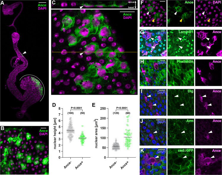
Fig 1. An Ance+ enterocyte subpopulation exhibits unique features.
(A) Cells in R2 (arrowhead) and R4 (white line) express Ance. (B) An enterocyte subpopulation displays Ance protein. (C) Nuclei of Ance+ cells (arrowhead) are flattened and lie in close apposition to the basal, visceral muscle. (D-E) Quantification of nuclear height (D) and nuclear area (E). (F) Some Ance+ enterocytes show weak (white arrowhead) or absent (yellow arrowhead) genome staining by DAPI. (G) Immunostaining of nuclear LaminB1 is reduced in Ance+ enterocytes (arrowheads). (H) Ance+ enterocytes decrease F-actin indicated by the absence of phalloidin staining. (I-K) Ance+ enterocytes exhibit less cell adhesion components. Immunostaining for Dlg labeling septate junctions (I) and β-catenin/Arm labeling adherens junctions (J) was reduced in Ance+ cells (arrowheads). Likewise, cadherin GFP knock-in flies demonstrate less adherens junctions in Ance+ enterocytes (arrowhead) (K). Statistical significance was determined by using a two-tailed unpaired t test (D and E). S1 Data provides the source data used for all graphs and statistical analyses. Scale bars, 200 μm (A), 20 μm (B-C, F-K). Arm, Armadillo; Dlg, Discs large.

