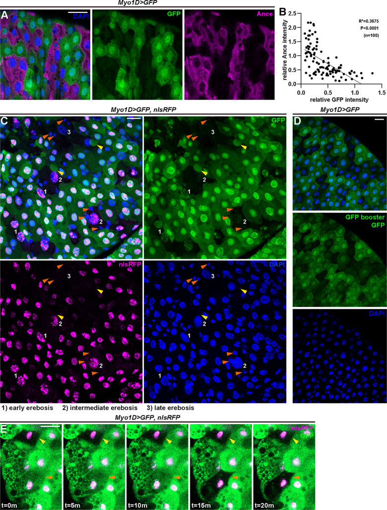
Fig 2. Ance+ enterocytes undergo erebosis.
(A) Ance+ immunostaining displays an inverse relationship to Myo1D-driven GFP fluorescence. Erebotic cells are labeled by Ance or loss of GFP signals. (B) A correlation analysis of median fluorescence intensity of Ance and GFP in the cell cytoplasm indicates that enterocytes exhibiting high Ance signals show low GFP signals and vice versa. Pearson’ correlation coefficient (R) was calculated: R = −0.6062, R2 = 0.3675, P < 0.0001. (C) Myo1D-driven GFP and nuclear RFP reveal a gradual process of erebosis. At early erebosis (1), cells lose cytoplasmic GFP but retain nuclear GFP and RFP. At intermediate erebosis (2), cells do not have signals of GFP but still retain nuclear RFP. At late erebosis (3), cells lose both GFP and RFP signals. Erebosis-induced protrusion can be observed in enterocytes near erebotic cells (yellow arrowheads). Small cells adjacent to erebotic cells are frequently visible (orange arrowheads). (D) GFP protein was not detectable by a GFP nanobody in erebotic enterocytes lacking Myo1D-driven GFP fluorescence. (E) Time-lapse imaging demonstrates that loss of GFP occurs in a live condition. The yellow arrowhead indicates a cell that had already undergone erebosis at the time of imaging. The orange arrowhead indicates a cell that lost cytoplasmic GFP during imaging. The loss of GFP occurred within 5 minutes. S1 Data provides the source data used for all graphs and statistical analyses. Scale bars, 20 μm.

