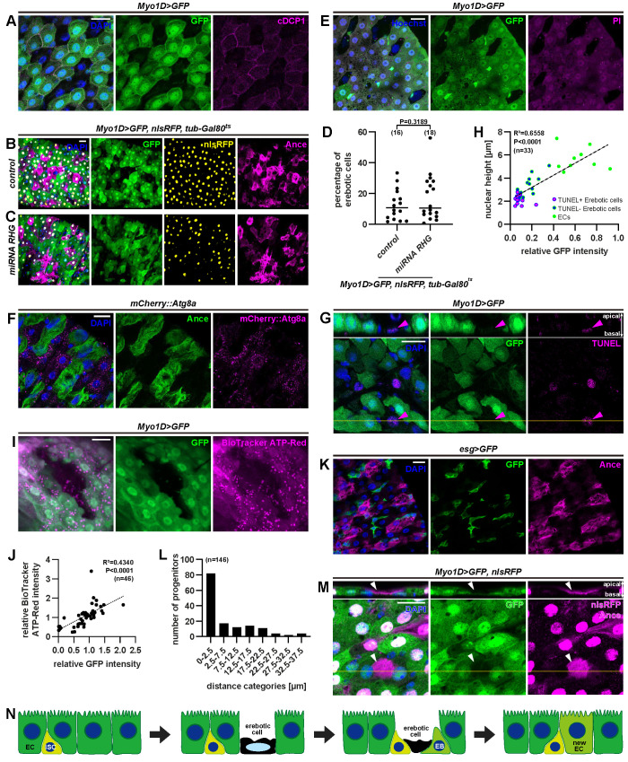
Fig 4. Erebosis is unprecedented cell death.
(A) Immunostaining for cleaved caspase 1 (cDCP1) does not show any increased signal in erebotic cells marked by absence of GFP fluorescence. (B, C) Erebotic cells indicated by absence of Myo1D-driven GFP and presence of Ance staining are visible even with suppression of apoptosis by expression of miRNA against rpr, hid, and grim. (D) Quantification of the percentage of erebotic cells with/without miRNA for rpr, hid, and grim. (E) Live imaging after PI feeding demonstrates that PI cannot enter erebotic cells. (F) Autophagosomes labeled by mCherry::Atg8a are reduced in erebotic (Ance+) cells. (G) Erebotic cells with a lower nuclear height and less GFP signals are labeled by TUNEL staining (arrowhead). (H) Correlation analysis of the nuclear height and nuclear GFP intensity demonstrates that cells at late erebosis (shorter nuclear height and weaker nuclear GFP signals) tend to be TUNEL positive. Pearson’ correlation coefficient (R) was calculated: R = 0.8098, R2 = 0.6558, P < 0.0001. (I) Erebotic cells have reduced amounts of cellular ATP labeled by BioTracker ATP-red, indicating their reduced metabolic activity. Note that the punctate signals of BioTracker ATP-red likely represent ATP in mitochondria. (J) Correlation analysis of ATP detected by BioTracker ATP-red and GFP intensity. Pearson’ correlation coefficient (R) was calculated: R = 0.6588, R2 = 0.4340, P < 0.0001. (K) Progenitor cells labeled by escargot-driven GFP (esg>GFP) are present in close proximity to Ance+ enterocytes. (L) Distance between erebotic cells and progenitor cells. (M) Immunostaining for Ance shows that a cytoplasmic legacy of an erebotic cell (arrowhead) is being pushed up by 2 enterocytes. The 2 enterocytes are considered to be relatively young because they have only GFP expression, not nlsRFP, and maturation of GFP is faster than of RFP. This captures a potential moment of the cell replacement. (N) Schematic of the erebosis process. Erebotic cells lose cytoskeleton, adhesion components, and organelles. Erebotic cells eventually become TUNEL positive, die, and are replaced by new enterocytes. Statistical significance was determined by using a two-tailed unpaired t test (D). S1 Data provides the source data used for all graphs and statistical analyses. Scale bars, 20 μm. EB, enteroblast; EC, enterocyte; ISC, intestinal stem cell; PI, propidium iodide.

