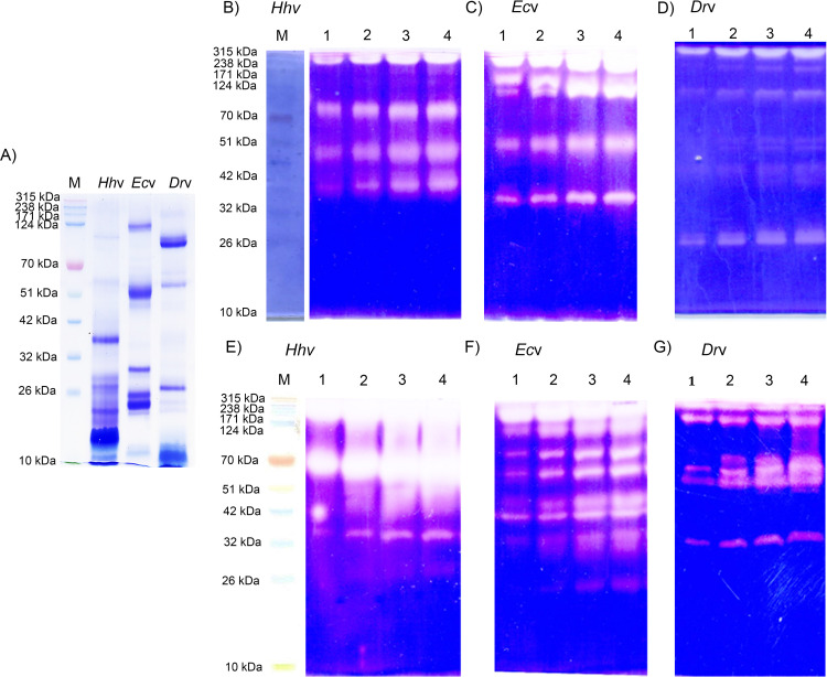
Fig 1. SDS-PAGE pattern, and zymography of H. hypnale, E. carinatus, and D. russelii venoms.
(A) SDS-PAGE (10%) banding pattern of venoms; Hhv, Ecv, and Drv (25 μg each) were used and analyzed under non-reducing condition. For zymography, the venoms Hhv, Ecv, and Drv were independently studied for proteolytic activity banding patterns in substrate gel assays. B-D and E-G respectively represent caseinolytic and gelatinolytic activities of three venoms. The substrates, casein, and gelatin (0.2%) were incorporated into respective gels (10%). The SDS-PAGE was performed under non-reduced condition. In lanes 1–4 in respective gels, different doses, 5, 10, 20, and 30 μg of venoms were used. M represents the molecular weight markers in kDa. The clear translucent zones against a blue background indicate the respective activities in gels. The images were captured by using HP Scanjet (Model-G2410).

