Figure 1.

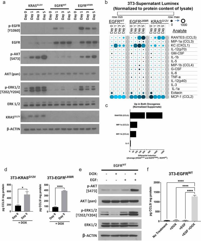
Oncogene signaling regulates cytokine production in premalignant cells. 3T3 cells were transduced with lentiviral constructs to express doxycyline (DOX) inducible wild-type EGFR, EGFRL858R, or KRASG12V. (a) Induced expression of oncogenic EGFRL858R and KRASG12V corresponded with increased phosphorylation of AKT and ERK, which was not seen with induced expression of EGFRWT. (b) Luminex assays were used to assess cytokines and chemokines produced after induction of EGFRWT, EGFRL858R, and KRASG12V. Data normalized to protein content of lysate (pg/mg) are visualized with a heatmap displaying levels of secreted protein for each cell line with increasing time post DOX treatment. Row Z-scores were calculated across all rows to show change in analyte and plotted, shown in color legend (row min-max). Value in picogram (pg) per milligram (mg) shown by circles small (0) to large (1000). (c) The analytes increased in both oncogene expressing cell lines are shown (see Supplemental Methods). (d) Secretion of CCL5 increased with 5 days of DOX treatment in 3T3 expressing KRASG12V and EGFRL858R as quantified by ELISA with DOX treatment day 0 (no treatment) and day 5. Induced expression of EGFRWT with the addition of 30 ng/mL EGF led to increased phosphorylation of AKT and ERK (e) and increased secretion of CCL5 (f). ELISA data show n = 3, with bars representing mean ± SEM, significance was determined by a Student’s T-test or an ANOVA with a post-hoc Dunnett’s test. *P < .05, **P ≤ 0.01, ***P ≤ 0.001, ****P ≤ 0.0001
