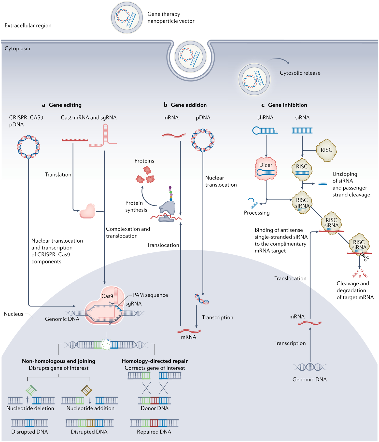
Fig. 1 |. Schematic pathways of three gene therapy avenues.

a | Gene editing. CRISPR–Cas9 coding DNA plasmid (pDNA) or Cas9 mRNA and single guide RNA (sgRNA) that contains a targeting sequence are delivered to the cell and enter the cytoplasm. pDNA translocates to the nucleus followed by transcription to Cas9 mRNA and sgRNA. Following translocation to the cytosol, Cas9 mRNA is translated to Cas9 protein and complexes sgRNA. Thereafter, Cas9–sgRNA complex translocates to the nucleus, where it binds the genomic DNA target sequence containing a proto-spacer adjacent motif (PAM) sequence at its 3’ end. Specific binding causes double-stranded DNA breakage resulting in non-homologous end joining or homology-directed repair, leading to mutations or changes within the gene sequence, respectively. b | Gene addition. After entering the cytoplasm, pDNA carrying the desired gene sequence translocates to the nucleus and undergoes transcription to mRNA. Next, transcripted or external mRNA is translated by the ribosome, promoting desired protein synthesis. c | Gene inhibition. After entering the cytoplasm, short hairpin RNA (shRNA) is detected and processed by Dicer protein to generate small interfering RNA (siRNA) later loaded onto RNA-induced silencing complex (RISC). Subsequently, double-stranded siRNA is unzipped, and passenger strand is cleaved. Antisense strand guides RISC towards target mRNA and aligns with it, thus leading to mRNA cleavage and degradation.
