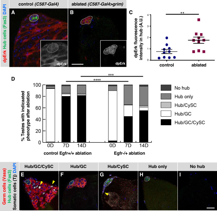
Figure 4. EGFR signaling is important for testis recovery from CySC ablation.
(A–B) Single confocal sections through the apex of testes immunostained for Fas3 (hub cell membranes, green), dpERK (EGFR pathway activation, red), and counterstained with DAPI (nuclei, blue). Hubs are outlined in white. Insets show the red channel alone, in white and enlarged. In control C587-Gal4, Gal80ts testes (A), dpERK levels are high in cyst lineage cells (indicating high levels of EGFR pathway activation) but low in hub cells. In C587-Gal4, Gal80ts > UAS grim testes (B), 2 days after genetic ablation of all CySCs and early cyst cells, dpERK levels are high in hub cells. Scale bar in B (for A-B) is 20 μm. (C) Quantification of dpERK levels in the hub in control (A) and ablated (B) testes. dpErk levels in the hub are significantly higher in ablated testes than in control testes. A.U., arbitrary units. Black bars indicate the mean and standard error. Unpaired t test, **p < 0.01. (D) Bar graph showing the distribution of testis phenotypes in control C587-Gal4, Gal80ts > UAS grim, Egfr+/+ flies ("control Egfr+/+ ablation") and in C587-Gal4, Gal80ts > UAS grim, Egfr-/+ flies ("EGFR-/+ ablation") at 0, 7, or 14 days after genetic ablation of CySCs and early cyst cells. After ablation (0 days), in both control and Egfr-/+ flies, most testes lack all CySCs and early cyst cells but retain a hub and germ cells (GC) as expected (white bars). At 7 and 14 days after ablation, fewer testes have regained CySCs and early cyst cells (black bars) in Egfr-/+ flies than in control flies and there is a significant difference in phenotype distribution. Chi square test, ***p < 0.001, ****p < 0.0001. (E–I) Single confocal sections through the apex of testes at 7 days after ablation, immunostained for Vasa (germ cells, red), Fas3 (hub cell membranes, green), and Tj (somatic cell nuclei, white), and counterstained with DAPI (nuclei, blue), to illustrate the phenotypes listed in (D). Testes that recover CySCs and early cyst lineage cells have Tj-positive nuclei outside the hub (yellow arrowheads); most also contain germ cells (E) but a few contain just a hub and cyst cells (G). Testes that fail to recover CySCs and early cyst lineage cells can retain a hub and germ cells (F) or just a hub (H) or no hub or germ cells (I). Hubs are outlined in white. Scale bar in I (for E-I) is 20 μm.

