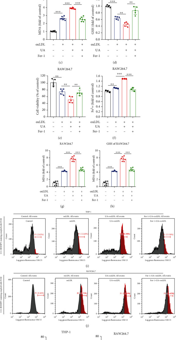
Figure 4.

HUA-induced ferroptosis is a key regulatory promoting foam cell formation. THP-1 and RAW264.7 cells were treated with oxLDL (100 μg/ml) or coincubated with UA (15 mg/dl) with or without Fer-1 (2 μM) for 24 h. (a and e) Cell viability was assayed by using a CCK-8 kit. (b and f) The accumulation of Fe2+ was measured by an iron detection assay. (c and g) Lipid formation was measured by MDA assay. (d and h) Relative GSH level was detected by using an assay kit. (i and j) C11-BODIPY staining coupled with FCM was used to assess lipid ROS levels. (k and l) Quantification of lipid ROS levels. Data are means ± SD, n = 3 − 7. FCM: flow cytometry; Fer-1: ferrostatin-1; GSH: glutathione; HUA: high level of uric acid; MDA: malondialdehyde; oxLDL: oxidized low-density lipoprotein; ROS: reactive oxygen species; UA: uric acid. ∗∗P < 0.01 and ∗∗∗P < 0.001.
