
Fig. 5. Transient expression of the 4F using TNNT2-4Fpolycistronic-NIL improves cardiac function and reduces scar size in rats after I/R.
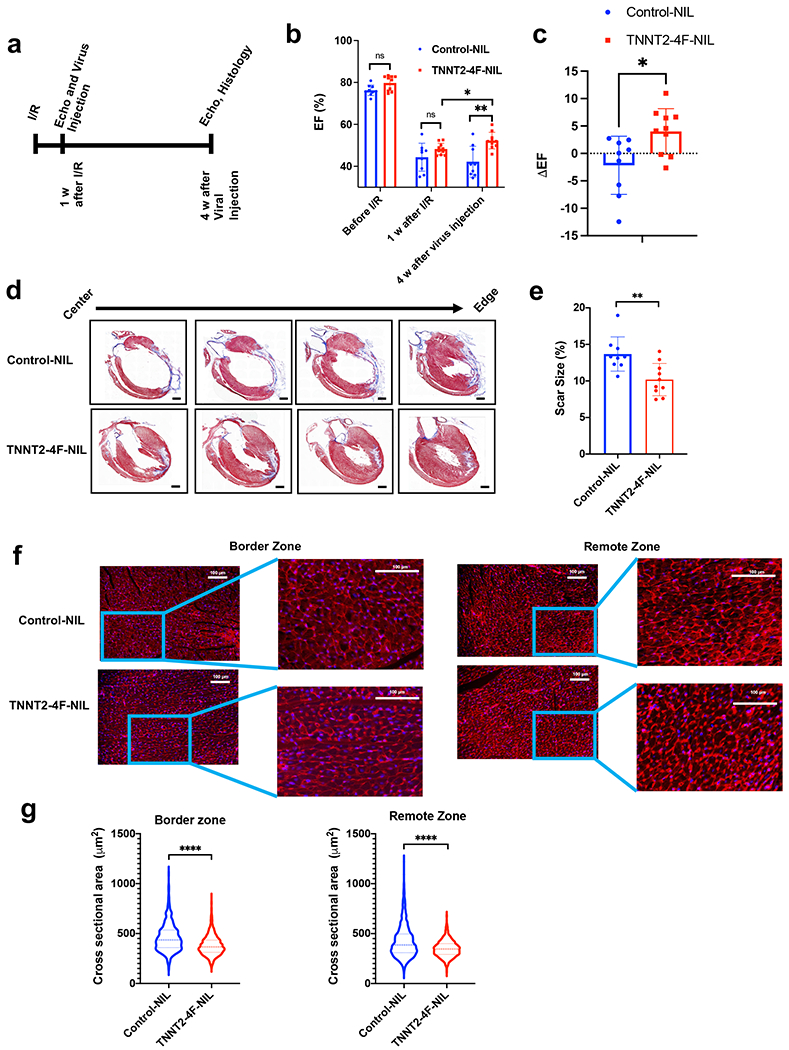
(a) schematic diagram of the experimental design. (b) Ejection fraction (EF), as assessed by echocardiography before Ischemia/reperfusion (I/R), one week after I/R (before viral treatment), and four weeks after viral treatment. (n=9-10 rats per group, *p<0.05, **p<0.01 compared to GFP-NIL control group, error bars indicate S.D.). (c) Quantification of the change in ejection fraction between before and after treatment for each individual rat (n=9-10 rats per group, *p<0.05, compared to GFP-NIL control group, error bars indicate S.D.). (d) Representative images of rat hearts were stained with Masson’s trichrome stain (healthy myocardium stains red and fibrotic tissue stains blue) at the end of the experiment (scale bar=2mm). (e) The scar size quantification as a percentage of total heart tissue (n=9-10 rats per group, 20-25 heart sections per animal, **p<0.01 compared to the control group, error bars indicate S.D.). (f) Representative images of rat heart at the border zone (right panel) or a remote zone (left panel) stained against wheat germ agglutinin, WGA (red), and nuclear DAPI (blue) (scale bar=100 µm). (g) Quantification of the cross-sectional area of cardiomyocytes at border zone (left) or a remote zone (right) (n=1300-1500 cells from each group, 20-25 heart sections per animal ****p<0.0001 compared to control group, error bars indicate S.D.).