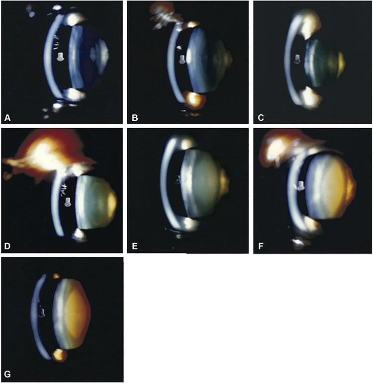
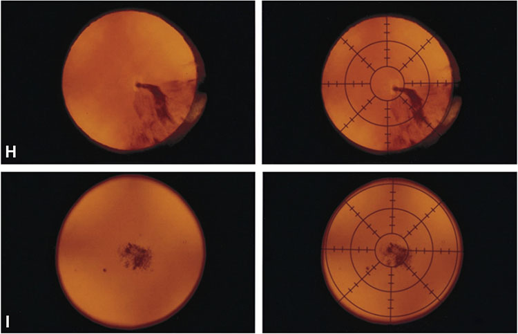
Figure 1. Reading center grading system for age-related cataract.
A-G, Nuclear cataract grading by comparison of 45-degree slit-lamp photograph with 7 standard photographs: 1 (no opacity) to 7 (extremely severe opacity). A—G, Standard photographs 1 through 7. H, Cortical cataract grading by percentage area involvement of the central 2 circles of the grid (5-mm diameter circular area) on retroillumination photograph. Left: Retroillumination photograph of cortical opacity. Right: Retroillumination photograph of cortical opacity with overlying grid. The cortical opacity occupies 22% of the central 2 circles of the grid. I, Posterior subcapsular cataract grading by percentage area involvement of the central 2 circles of the grid (5-mm diameter circular area) on retroillumination photograph. Left: Retroillumination photograph of posterior subcapsular opacity. Right: Retroillumination photograph of posterior subcapsular opacity with overlying grid. The posterior subcapsular opacity occupies 15% of the central 2 circles of the grid.


