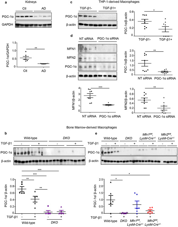
Figure 2. Mitochondrial biogenesis is impaired during experimental kidney fibrosis and PGC-1α regulates expressions of MFN1 and MFN2 in macrophages.
Western blot and densitometry analysis for the expression of PGC-1α in a) kidney tissue lysates from wild-type mice fed with control (Ctl) or adenine diet (AD) (n = 6 per group) for 28 days, normalized to GAPDH; b) bone marrow-derived macrophages (BMDM) isolated from wild-type, Mfn1/Mfn2 double knockout (DKO) mice treated with TGF-β1 (5 ng/ml) for 48 hours (n = 5 per group), normalized to β-actin (n = 5 per group), c) THP-1-derived human macrophages mice treated with TGF-β1 (5 ng/ml) for 48 hours (n = 8 per group), d) MFN1, MFN2 expression in THP-1-derived human macrophages transfected with PGC-1α siRNA or non-targeting (NT) control siRNA (n = 8 per group), normalized to β-actin. e) PGC-1α expression in BMDM isolated from wild-type, DKO, Mfn1-, Mfn2- deficient mice treated with TGF-β1 (5 ng/ml) for 48 hours (n = 6 per group), normalized to β-actin Data are mean ± SEM representative of 3 independent experiments. *P < 0.05, **P < 0.01, ***P < 0.001, analyzed by student’s unpaired 1-tailed t-test (a, c, d) or one-way ANOVA followed by Newman-Keuls post-hoc test (b, e).

