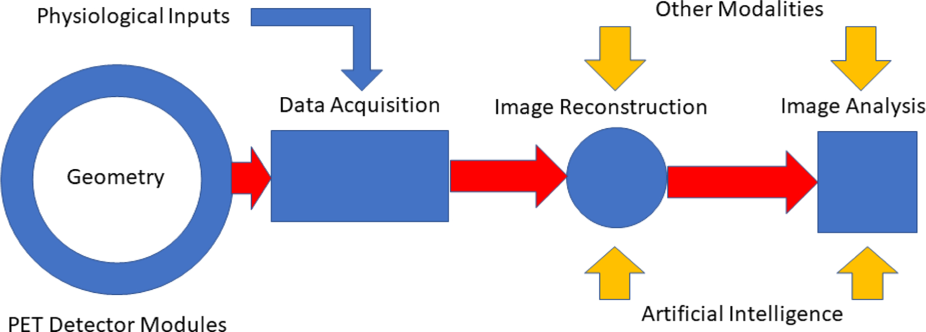
Figure 1.

Schematic illustration of an idealized small animal PET imaging system. Relatively low temporal resolution physiological inputs include chest cuff pressure signals, ECG-R wave markers and stimulus gating signals. Compact, high density digital PET detector modules surrounding the full length of the imaging subject provide high temporal resolution LIST mode input of spatial, timing and energy signals from time coincident gamma ray events in the detector array. During image reconstruction these signals are transformed into PET images corrected for a host of confounding effects, e.g., attenuation, scatter, positron range, using physical models of these processes and, more recently, by artificial intelligence methods that achieve some of these same ends. Combination of these images with those from other modalities acquired serially or simultaneously provide complementary information about target structure and function.
