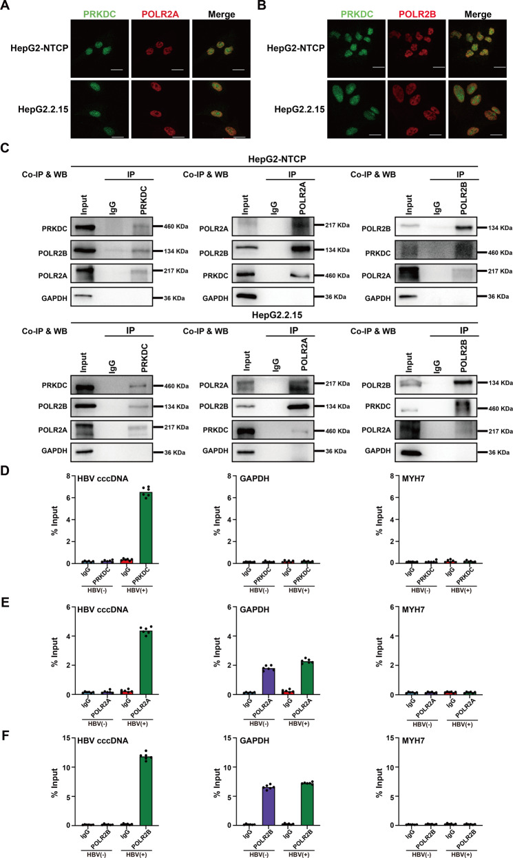
Fig. 5. PRKDC interacts with POLR2A and POLR2B, and associates with cccDNA.
A, B Immunofluorescent staining demonstrates partial colocalization of PRKDC (green) and POLR2A (A) or POLR2B (B) in HBV-infected HepG2-NTCP and HepG2.2.15 cells. Scale bar, 10 μm. C Endogenous co-immunoprecipitation with PRKDC (left), POLR2A (middle), and POLR2B (right) was carried out in HBV-infected HepG2-NTCP and HepG2.2.15 cells using indicated antibodies and blotted with specific antibodies. D–F Cross-linked chromatin from HBV-infected or non-HBV-infected HepG2-NTCP cells was immunoprecipitated with PRKDC (D), POLR2A (E), and POLR2B (F), and the corresponding IgG was used as a control, followed by PCR quantification of HBV cccDNA and the promoter of GAPDH and MYH7 using specific primers. The results are displayed as the percentage of input.

