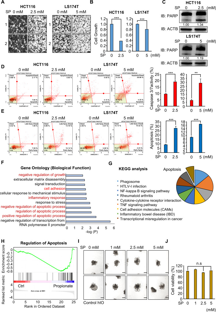
Fig. 1. Propionate suppresses the cell growth of HCT116 and LS174T cells via cell apoptosis.
A, B Cell growth assay after sodium propionate treatment for 48 h. HCT116 and LS174T cells were fixed in 100% methanol and stained with crystal violet solution. Scale bar, 200 μm (A). Cells were incubated for 5 min at 37 °C after adding CCK-8 solution. The intensity of cell growth was measured using a microplate reader (450 nm). Mean ± SD of three independent experiments. p values were calculated using Student’s t-test (***p < 0.001) (B). C Western blot analysis after sodium propionate treatment using anti-PARP. ACTB was used as the internal control in HCT116 and LS174T cells. The signal intensities were quantified using ImageJ software. D FACS analysis using Muse Caspase 3/7 working solution was performed after sodium propionate treatment. The upper right panel indicates the apoptotic and dead cell proportions (left). Quantification of caspase 3/7 activity. Mean ± SD of three independent experiments. p values were calculated using Student’s t-test (***p < 0.001, **p < 0.01) (right). E FACS analysis of Annexin V staining was performed after sodium propionate treatment. The lower right and upper right quadrants indicate early apoptosis and late apoptosis (left). Quantification of apoptosis. Mean ± SD of three independent experiments. p values were calculated using Student’s t-test (***p < 0.001) (right). F DAVID-based GO analysis of RNA-seq results of the up- and downregulated genes among the 872 genes. G DAVID-based KEGG pathway analysis of 872 genes. The enriched terms are shown. H GSEA using 872 genes. I Representative images of the morphology of hIOs after treatment with varying concentrations of propionate. Scale bar, 500 μm. J Cell viability assessed by the CCK assay in hIO treated with the indicated concentrations of propionate. n = 3 per group. Mean ± SD of three independent experiments. p values were calculated using Student’s t-test (n.s. nonsignificant).

