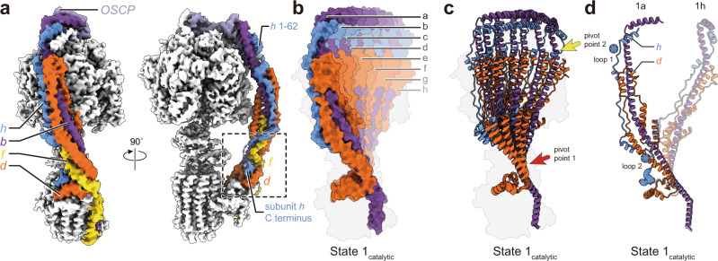
Fig. 3. Deformation of the peripheral stalk of ATP synthase while under strain during ATP hydrolysis.
a Structure of the peripheral stalk shows that subunit h bridges F1 and FO. b Overlay of the eight State 1catalytic atomic models, produced by molecular dynamics flexible fitting of the atomic model of resting ATP synthase into the eight 3D maps of State 1catalytic, shows a large deformation of the peripheral stalk. c The peripheral stalk bends at two pivot points near F1 (yellow arrow) and near FO (red arrow). d Unstructured regions in subunit h (blue asterisks) allow it to withstand the bending of subunit d.

