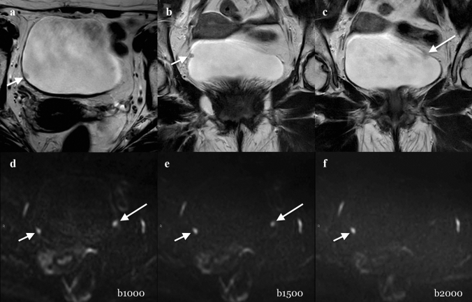
Fig. 4.
Example of a not muscle-invasive BC classified incorrectly. A 73-year-old man with hematuria and two polyps, documented after flexible cystoscopy, underwent MRI before primary TURB. Axial (a) and coronal (b, c) T2W imaging (T2) showed a small (4 mm) polypoid lesion on the right wall of the bladder (short arrow in a and b). The lesion was well detected by the three readers regardless the image set and was scored as VI-RADS 1 (short arrow in d, e, and f). Another slightly visible small (4 mm) non-muscular invasive lesion (VI-RADS 1) on the left wall of the bladder was suspected on T2 images (long arrow in c). However, it was definitely detected by the three readers only when reading b1000 and b1500 images (long arrow in d and e), but not on b2000 images. T stage after TURB was LG-T1 (TURB). After four weeks, Re-TURB was performed and it confirmed the absence of residual tumor. DWI = diffusion-weighted imaging; HG = high grade; MRI = magnetic resonance imaging; T2W = T2 weighted; TURB = transurethral resection of the bladder; VI-RADS = Vesical Imaging-Reporting and Data System

