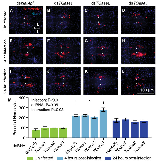
Figure 2.
RNAi-based knockdown of TGase3 increases the number of periostial hemocytes during the early stages of GFP-E. coli infection. (A–L) Each fluorescence image shows the tergum of a single abdominal segment with periostial hemocytes (CM-DiI; red) surrounding the ostia (asterisks) in uninfected mosquitoes (A–D), and mosquitoes at 4 (E–H) or 24 h (I–L) post-infection. Prior to treatment, mosquitoes had been injected with dsbla(ApR) (A, E, I), dsTGase1 (B, F, J), dsTGase2 (C, G, K) or dsTGase3 (D, H, L). Nuclei were stained blue with Hoechst 33342. A, anterior; P, posterior; L, lateral. (M) Graph shows the average number of periostial hemocytes in dsbla(ApR)-, dsTGase1-, dsTGase2- and dsTGase3-injected mosquitoes that were uninfected or had been infected with GFP-E. coli for 4 or 24 h. Data were analyzed by two-way ANOVA followed by Dunnett’s post-hoc test, using dsbla(ApR) mosquitoes as the reference. Infection P indicates whether infection has an effect irrespective of dsRNA treatment, dsRNA P indicates whether dsRNA treatment has an effect irrespective of infection, and interaction P indicates whether the effect of infection changes with dsRNA treatment. Column heights mark the means, whiskers show the standard error of the mean (S.E.M.), and the asterisk indicates post-hoc P < 0.05.

